Enzyme intermediates captured "on the fly" by mix-and-inject serial crystallography.
Olmos, J.L., Pandey, S., Martin-Garcia, J.M., Calvey, G., Katz, A., Knoska, J., Kupitz, C., Hunter, M.S., Liang, M., Oberthuer, D., Yefanov, O., Wiedorn, M., Heyman, M., Holl, M., Pande, K., Barty, A., Miller, M.D., Stern, S., Roy-Chowdhury, S., Coe, J., Nagaratnam, N., Zook, J., Verburgt, J., Norwood, T., Poudyal, I., Xu, D., Koglin, J., Seaberg, M.H., Zhao, Y., Bajt, S., Grant, T., Mariani, V., Nelson, G., Subramanian, G., Bae, E., Fromme, R., Fung, R., Schwander, P., Frank, M., White, T.A., Weierstall, U., Zatsepin, N., Spence, J., Fromme, P., Chapman, H.N., Pollack, L., Tremblay, L., Ourmazd, A., Phillips, G.N., Schmidt, M.(2018) BMC Biol 16: 59-59
- PubMed: 29848358
- DOI: https://doi.org/10.1186/s12915-018-0524-5
- Primary Citation of Related Structures:
6B5X, 6B5Y, 6B68, 6B69, 6B6A, 6B6B, 6B6C, 6B6D, 6B6E, 6B6F - PubMed Abstract:
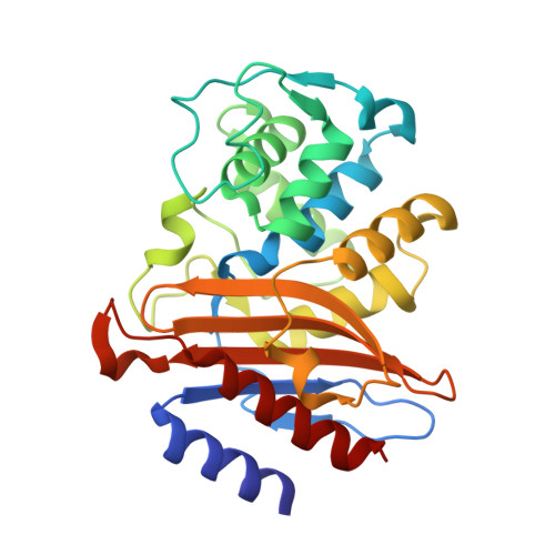
Ever since the first atomic structure of an enzyme was solved, the discovery of the mechanism and dynamics of reactions catalyzed by biomolecules has been the key goal for the understanding of the molecular processes that drive life on earth. Despite a large number of successful methods for trapping reaction intermediates, the direct observation of an ongoing reaction has been possible only in rare and exceptional cases. Here, we demonstrate a general method for capturing enzyme catalysis "in action" by mix-and-inject serial crystallography (MISC). Specifically, we follow the catalytic reaction of the Mycobacterium tuberculosis β-lactamase with the third-generation antibiotic ceftriaxone by time-resolved serial femtosecond crystallography. The results reveal, in near atomic detail, antibiotic cleavage and inactivation from 30 ms to 2 s. MISC is a versatile and generally applicable method to investigate reactions of biological macromolecules, some of which are of immense biological significance and might be, in addition, important targets for structure-based drug design. With megahertz X-ray pulse rates expected at the Linac Coherent Light Source II and the European X-ray free-electron laser, multiple, finely spaced time delays can be collected rapidly, allowing a comprehensive description of biomolecular reactions in terms of structure and kinetics from the same set of X-ray data.
Organizational Affiliation:
Department of BioSciences, Rice University, 6100 Main Street, Houston, TX, 77005, USA.