6AR4

Crystal structure of PICK1 in complex with the small molecule inhibitor 1o


Experimental Data Snapshot

  • Method: X-RAY DIFFRACTION
  • Resolution: 1.69 Å
  • R-Value Free: 0.190 
  • R-Value Work: 0.157 
  • R-Value Observed: 0.159 

wwPDB Validation   3D Report Full Report


Ligand Structure Quality Assessment 


This is version 1.2 of the entry. See complete history


Literature

Potent PDZ-Domain PICK1 Inhibitors that Modulate Amyloid Beta-Mediated Synaptic Dysfunction.

Lin, E.Y.S.Silvian, L.F.Marcotte, D.J.Banos, C.C.Jow, F.Chan, T.R.Arduini, R.M.Qian, F.Baker, D.P.Bergeron, C.Hession, C.A.Huganir, R.L.Borenstein, C.F.Enyedy, I.Zou, J.Rohde, E.Wittmann, M.Kumaravel, G.Rhodes, K.J.Scannevin, R.H.Dunah, A.W.Guckian, K.M.

(2018) Sci Rep 8: 13438-13438

  • DOI: https://doi.org/10.1038/s41598-018-31680-3
  • Primary Citation of Related Structures:  
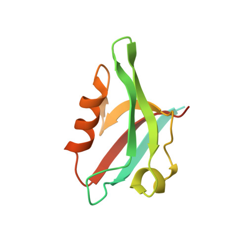
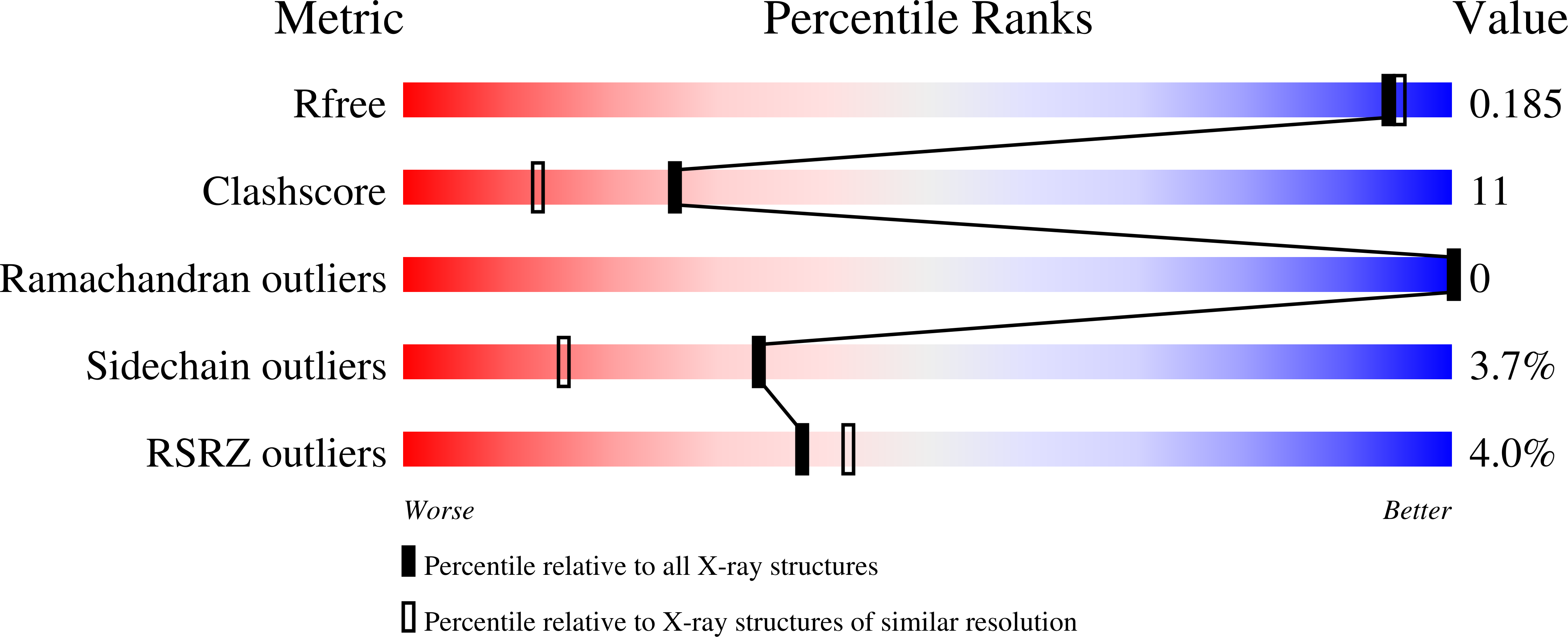
    6AR4

  • PubMed Abstract: 

    Protein interacting with C kinase (PICK1) is a scaffolding protein that is present in dendritic spines and interacts with a wide array of proteins through its PDZ domain. The best understood function of PICK1 is regulation of trafficking of AMPA receptors at neuronal synapses via its specific interaction with the AMPA GluA2 subunit. Disrupting the PICK1-GluA2 interaction has been shown to alter synaptic plasticity, a molecular mechanism of learning and memory. Lack of potent, selective inhibitors of the PICK1 PDZ domain has hindered efforts at exploring the PICK1-GluA2 interaction as a therapeutic target for neurological diseases. Here, we report the discovery of PICK1 small molecule inhibitors using a structure-based drug design strategy. The inhibitors stabilized surface GluA2, reduced Aβ-induced rise in intracellular calcium concentrations in cultured neurons, and blocked long term depression in brain slices. These findings demonstrate that it is possible to identify potent, selective PICK1-GluA2 inhibitors which may prove useful for treatment of neurodegenerative disorders.


  • Organizational Affiliation

    Biotherapeutics and Medicinal Sciences, Biogen Inc, Cambridge, Massachusetts, USA.


Macromolecules
Find similar proteins by:  (by identity cutoff)  |  3D Structure
Entity ID: 1
MoleculeChains Sequence LengthOrganismDetailsImage
PRKCA-binding protein
A, B
125Homo sapiensMutation(s): 0 
Gene Names: PICK1PRKCABP
UniProt & NIH Common Fund Data Resources
Find proteins for Q9NRD5 (Homo sapiens)
Explore Q9NRD5 
Go to UniProtKB:  Q9NRD5
PHAROS:  Q9NRD5
GTEx:  ENSG00000100151 
Entity Groups  
Sequence Clusters30% Identity50% Identity70% Identity90% Identity95% Identity100% Identity
UniProt GroupQ9NRD5
Sequence Annotations
Expand
  • Reference Sequence
Small Molecules
Ligands 1 Unique
IDChains Name / Formula / InChI Key2D Diagram3D Interactions
BQA
Query on BQA

Download Ideal Coordinates CCD File 
C [auth A],
D [auth B]
N-[4-(4-bromophenyl)-1-{[2-(trifluoromethyl)phenyl]methyl}piperidine-4-carbonyl]-3-cyclopropyl-L-alanine
C26 H28 Br F3 N2 O3
XTCYTRMSAWHHGP-QFIPXVFZSA-N
Binding Affinity Annotations 
IDSourceBinding Affinity
BQA Binding MOAD:  6AR4 IC50: 573 (nM) from 1 assay(s)
Experimental Data & Validation

Experimental Data

  • Method: X-RAY DIFFRACTION
  • Resolution: 1.69 Å
  • R-Value Free: 0.190 
  • R-Value Work: 0.157 
  • R-Value Observed: 0.159 
  • Space Group: P 32
Unit Cell:
Length ( Å )Angle ( ˚ )
a = 54.311α = 90
b = 54.311β = 90
c = 77.633γ = 120
Software Package:
Software NamePurpose
Aimlessdata scaling
REFMACrefinement
PDB_EXTRACTdata extraction
MOLREPphasing

Structure Validation

View Full Validation Report


Ligand Structure Quality Assessment 


Entry History 

Deposition Data

  • Released Date: 2018-08-22 
  • Deposition Author(s): Marcotte, D.

Revision History  (Full details and data files)

  • Version 1.0: 2018-08-22
    Type: Initial release
  • Version 1.1: 2018-09-19
    Changes: Data collection, Database references, Structure summary
  • Version 1.2: 2023-10-04
    Changes: Data collection, Database references, Refinement description