6AA4

Crystal structure of MTH1 in complex with alpha-mangostin (cocktail No. 9)


Experimental Data Snapshot

  • Method: X-RAY DIFFRACTION
  • Resolution: 1.90 Å
  • R-Value Free: 0.244 
  • R-Value Work: 0.206 
  • R-Value Observed: 0.208 

wwPDB Validation   3D Report Full Report


Ligand Structure Quality Assessment 


This is version 1.1 of the entry. See complete history


Literature

Discovery of a new class of MTH1 inhibitor by X-ray crystallographic screening.

Yokoyama, T.Kitakami, R.Mizuguchi, M.

(2019) Eur J Med Chem 167: 153-160

  • DOI: https://doi.org/10.1016/j.ejmech.2019.02.011
  • Primary Citation of Related Structures:  
    6AA3, 6AA4, 6AA5

  • PubMed Abstract: 

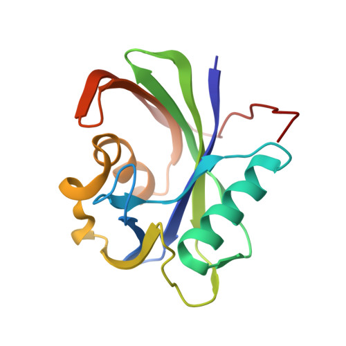
    MutT homologue 1 (MTH1) protects the nucleotide pool from oxidative stress by hydrolyzing oxidized nucleoside triphosphates and prevents their incorporation into DNA. Cancer cells are dependent on the MTH1 activity for survival due to the high-level of reactive oxygen species in cancer cells; therefore, MTH1 is considered to be a novel target for treatment of various cancers. Here, we show by X-ray crystallographic screening using an in-house cocktail library that α-mangostin, a natural xanthone from mangosteen pericarp, binds to the active site of MTH1. A subsequent inhibition assay revealed that 3-isomangostin, a cyclized derivative of α-mangostin, was the most potent MTH1 inhibitor, with an IC 50 value of 0.052 μM. Detailed structural analyses of the MTH1-3-isomangostin complex showed the novel binding mode of 3-isomangostin. Our results demonstrate that X-ray crystallographic screening is useful for the lead discovery for MTH1, and suggest that 3-isomangostin would be an attractive chemical tool for the development of anticancer agents.


  • Organizational Affiliation

    Faculty of Pharmaceutical Sciences, University of Toyama, 2630 Sugitani, Toyama, 930-0914, Japan. Electronic address: tyokoya3@pha.u-toyama.ac.jp.


Macromolecules
Find similar proteins by:  (by identity cutoff)  |  3D Structure
Entity ID: 1
MoleculeChains Sequence LengthOrganismDetailsImage
7,8-dihydro-8-oxoguanine triphosphatase163Homo sapiensMutation(s): 0 
Gene Names: NUDT1MTH1
EC: 3.6.1.55 (PDB Primary Data), 3.6.1.56 (PDB Primary Data)
UniProt & NIH Common Fund Data Resources
Find proteins for P36639 (Homo sapiens)
Explore P36639 
Go to UniProtKB:  P36639
PHAROS:  P36639
GTEx:  ENSG00000106268 
Entity Groups  
Sequence Clusters30% Identity50% Identity70% Identity90% Identity95% Identity100% Identity
UniProt GroupP36639
Sequence Annotations
Expand
  • Reference Sequence
Small Molecules
Binding Affinity Annotations 
IDSourceBinding Affinity
MKS Binding MOAD:  6AA4 IC50: 470 (nM) from 1 assay(s)
Experimental Data & Validation

Experimental Data

  • Method: X-RAY DIFFRACTION
  • Resolution: 1.90 Å
  • R-Value Free: 0.244 
  • R-Value Work: 0.206 
  • R-Value Observed: 0.208 
  • Space Group: I 4 2 2
Unit Cell:
Length ( Å )Angle ( ˚ )
a = 66.796α = 90
b = 66.796β = 90
c = 140.598γ = 90
Software Package:
Software NamePurpose
PHENIXrefinement
XDSdata reduction
XSCALEdata scaling
PHENIXphasing

Structure Validation

View Full Validation Report


Ligand Structure Quality Assessment 


Entry History 

Deposition Data

Revision History  (Full details and data files)

  • Version 1.0: 2019-03-06
    Type: Initial release
  • Version 1.1: 2023-11-22
    Changes: Data collection, Database references, Derived calculations, Refinement description