6A9J

Crystal structure of the PE-bound N-terminal domain of Atg2


Experimental Data Snapshot

  • Method: X-RAY DIFFRACTION
  • Resolution: 2.70 Å
  • R-Value Free: 0.268 
  • R-Value Work: 0.251 
  • R-Value Observed: 0.256 

wwPDB Validation   3D Report Full Report


Ligand Structure Quality Assessment 


This is version 2.1 of the entry. See complete history


Literature

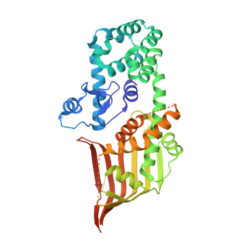
Atg2 mediates direct lipid transfer between membranes for autophagosome formation.

Osawa, T.Kotani, T.Kawaoka, T.Hirata, E.Suzuki, K.Nakatogawa, H.Ohsumi, Y.Noda, N.N.

(2019) Nat Struct Mol Biol 26: 281-288

  • DOI: https://doi.org/10.1038/s41594-019-0203-4
  • Primary Citation of Related Structures:  
    6A9E, 6A9J

  • PubMed Abstract: 

    A key event in autophagy is autophagosome formation, whereby the newly synthesized isolation membrane (IM) expands to form a complete autophagosome using endomembrane-derived lipids. Atg2 physically links the edge of the expanding IM with the endoplasmic reticulum (ER), a role that is essential for autophagosome formation. However, the molecular function of Atg2 during ER-IM contact remains unclear, as does the mechanism of lipid delivery to the IM. Here we show that the conserved amino-terminal region of Schizosaccharomyces pombe Atg2 includes a lipid-transfer-protein-like hydrophobic cavity that accommodates phospholipid acyl chains. Atg2 bridges highly curved liposomes, thereby facilitating efficient phospholipid transfer in vitro, a function that is inhibited by mutations that impair autophagosome formation in vivo. These results suggest that Atg2 acts as a lipid-transfer protein that supplies phospholipids for autophagosome formation.


  • Organizational Affiliation

    Institute of Microbial Chemistry (BIKAKEN), Tokyo, Japan.


Macromolecules
Find similar proteins by:  (by identity cutoff)  |  3D Structure
Entity ID: 1
MoleculeChains Sequence LengthOrganismDetailsImage
Endolysin,Autophagy-related protein 2
A, B
389Enterobacteria phage RB59Schizosaccharomyces pombe 972h-
This entity is chimeric
Mutation(s): 3 
Gene Names: eRB59_126atg2mug36SPBC31E1.01cSPBC660.18c
EC: 3.2.1.17
UniProt
Find proteins for O94649 (Schizosaccharomyces pombe (strain 972 / ATCC 24843))
Explore O94649 
Go to UniProtKB:  O94649
Find proteins for P00720 (Enterobacteria phage T4)
Explore P00720 
Go to UniProtKB:  P00720
Entity Groups  
Sequence Clusters30% Identity50% Identity70% Identity90% Identity95% Identity100% Identity
UniProt GroupsP00720O94649
Sequence Annotations
Expand
  • Reference Sequence
Small Molecules
Ligands 1 Unique
IDChains Name / Formula / InChI Key2D Diagram3D Interactions
PEE
Query on PEE

Download Ideal Coordinates CCD File 
C [auth A],
D [auth B]
1,2-dioleoyl-sn-glycero-3-phosphoethanolamine
C41 H78 N O8 P
MWRBNPKJOOWZPW-NYVOMTAGSA-N
Experimental Data & Validation

Experimental Data

  • Method: X-RAY DIFFRACTION
  • Resolution: 2.70 Å
  • R-Value Free: 0.268 
  • R-Value Work: 0.251 
  • R-Value Observed: 0.256 
  • Space Group: P 1 21 1
Unit Cell:
Length ( Å )Angle ( ˚ )
a = 86.657α = 90
b = 62.861β = 91.89
c = 86.662γ = 90
Software Package:
Software NamePurpose
PHENIXrefinement
HKL-2000data reduction
HKL-2000data scaling
PHENIXphasing

Structure Validation

View Full Validation Report


Ligand Structure Quality Assessment 


Entry History 

Deposition Data

Revision History  (Full details and data files)

  • Version 1.0: 2019-03-20
    Type: Initial release
  • Version 1.1: 2019-03-27
    Changes: Data collection, Database references
  • Version 1.2: 2019-04-10
    Changes: Data collection, Database references
  • Version 1.3: 2019-04-17
    Changes: Data collection, Database references
  • Version 2.0: 2021-10-06
    Changes: Atomic model, Data collection, Database references, Derived calculations, Non-polymer description, Structure summary
  • Version 2.1: 2023-11-22
    Changes: Data collection, Refinement description