6A4U

The first crystal structure of crustacean ferritin that is a hybrid type of H and L ferritin


Experimental Data Snapshot

  • Method: X-RAY DIFFRACTION
  • Resolution: 1.16 Å
  • R-Value Free: 0.157 
  • R-Value Work: 0.138 
  • R-Value Observed: 0.139 

wwPDB Validation   3D Report Full Report


This is version 1.2 of the entry. See complete history


Literature

The first crystal structure of crustacean ferritin that is a hybrid type of H and L ferritin

Masuda, T.Zang, J.Zhao, G.Mikami, B.

(2018) Protein Sci 27: 1955-1960

  • DOI: https://doi.org/10.1002/pro.3495
  • Primary Citation of Related Structures:  
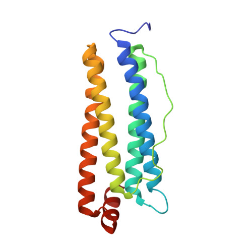
    6A4U

  • PubMed Abstract: 

    Ferritin, a ubiquitous iron storage protein, has a crucial role in innate immunity in arthropods, which have no adaptive immune system. Arthropods are thought to have two types of ferritin molecules: the secreted type and the cytosolic type. Here, we present the first crystal structure of ferritin from crustacean, kuruma prawn (Marsupenaeus japonicus), at 1.16 Å resolution. This shrimp ferritin (MjFer) is the cytosolic type, and its structure shows well-conserved ferritin fold composed of a 4-helix bundle that assembles into a cage-like 24-mer. The structure of MjFer was more similar to those of human and vertebrate ferritins than to that of the secreted-type arthropod ferritin from an insect. MjFer possesses both a ferroxidase site and a nucleation site, which are the main characteristics of vertebrate H and L chain ferritins, respectively. The first crystal structure of crustacean ferritin, MjFer, has exceptionally high quality that provides the detailed structural information of metal moving pathway in ferritin.


  • Organizational Affiliation

    Laboratory of Food Quality Design and Development, Division of Agronomy and Horticultural Science, Graduate School of Agriculture, Kyoto University, Kyoto, 611-0011, Japan.


Macromolecules
Find similar proteins by:  (by identity cutoff)  |  3D Structure
Entity ID: 1
MoleculeChains Sequence LengthOrganismDetailsImage
Ferritin
A, B, C, D, E
A, B, C, D, E, F
169Penaeus japonicusMutation(s): 1 
EC: 1.16.3.1
UniProt
Find proteins for T2B7E1 (Penaeus japonicus)
Explore T2B7E1 
Go to UniProtKB:  T2B7E1
Entity Groups  
Sequence Clusters30% Identity50% Identity70% Identity90% Identity95% Identity100% Identity
UniProt GroupT2B7E1
Sequence Annotations
Expand
  • Reference Sequence
Small Molecules
Ligands 4 Unique
IDChains Name / Formula / InChI Key2D Diagram3D Interactions
SO4
Query on SO4

Download Ideal Coordinates CCD File 
AA [auth B]
BA [auth B]
LB [auth E]
MB [auth E]
NA [auth C]
AA [auth B],
BA [auth B],
LB [auth E],
MB [auth E],
NA [auth C],
P [auth A],
Q [auth A],
R [auth A],
UB [auth F],
VA [auth D],
VB [auth F],
WA [auth D],
Z [auth B]
SULFATE ION
O4 S
QAOWNCQODCNURD-UHFFFAOYSA-L
EDO
Query on EDO

Download Ideal Coordinates CCD File 
AB [auth E]
BB [auth E]
CB [auth E]
DA [auth C]
DB [auth E]
AB [auth E],
BB [auth E],
CB [auth E],
DA [auth C],
DB [auth E],
EA [auth C],
EB [auth E],
FA [auth C],
FB [auth E],
G [auth A],
GA [auth C],
GB [auth E],
H [auth A],
HA [auth C],
HB [auth E],
I [auth A],
IA [auth C],
IB [auth E],
J [auth A],
JA [auth C],
JB [auth E],
K [auth A],
KA [auth C],
KB [auth E],
L [auth A],
LA [auth C],
M [auth A],
MA [auth C],
N [auth A],
NB [auth F],
O [auth A],
OB [auth F],
PB [auth F],
QA [auth D],
QB [auth F],
RA [auth D],
RB [auth F],
S [auth B],
SA [auth D],
SB [auth F],
T [auth B],
TA [auth D],
TB [auth F],
U [auth B],
UA [auth D],
V [auth B],
W [auth B],
X [auth B],
Y [auth B],
ZA [auth E]
1,2-ETHANEDIOL
C2 H6 O2
LYCAIKOWRPUZTN-UHFFFAOYSA-N
CL
Query on CL

Download Ideal Coordinates CCD File 
CA [auth B]CHLORIDE ION
Cl
VEXZGXHMUGYJMC-UHFFFAOYSA-M
MG
Query on MG

Download Ideal Coordinates CCD File 
OA [auth C],
PA [auth C],
WB [auth F],
XA [auth D],
YA [auth D]
MAGNESIUM ION
Mg
JLVVSXFLKOJNIY-UHFFFAOYSA-N
Experimental Data & Validation

Experimental Data

  • Method: X-RAY DIFFRACTION
  • Resolution: 1.16 Å
  • R-Value Free: 0.157 
  • R-Value Work: 0.138 
  • R-Value Observed: 0.139 
  • Space Group: I 4
Unit Cell:
Length ( Å )Angle ( ˚ )
a = 124.872α = 90
b = 124.872β = 90
c = 175.683γ = 90
Software Package:
Software NamePurpose
PHENIXrefinement
HKL-2000data reduction
HKL-2000data scaling
MOLREPphasing

Structure Validation

View Full Validation Report


Entry History & Funding Information

Deposition Data


Funding OrganizationLocationGrant Number
Japan Society for the Promotion of ScienceJapanJP 15K07574

Revision History  (Full details and data files)

  • Version 1.0: 2018-08-22
    Type: Initial release
  • Version 1.1: 2018-11-07
    Changes: Data collection, Database references
  • Version 1.2: 2023-11-22
    Changes: Data collection, Database references, Derived calculations, Refinement description