6A4S

Crystal structure of peptidase E with ordered active site loop from Salmonella enterica


Experimental Data Snapshot

  • Method: X-RAY DIFFRACTION
  • Resolution: 1.90 Å
  • R-Value Free: 0.211 
  • R-Value Work: 0.182 
  • R-Value Observed: 0.183 

wwPDB Validation   3D Report Full Report


This is version 1.1 of the entry. See complete history


Literature

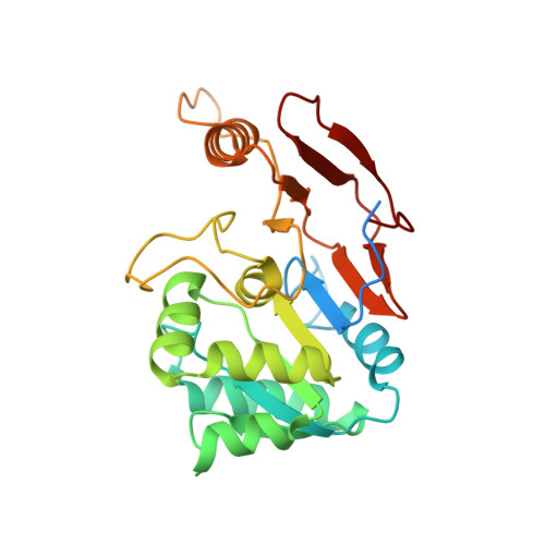
Structure of Asp-bound peptidase E from Salmonella enterica: Active site at dimer interface illuminates Asp recognition.

Yadav, P.Goyal, V.D.Gaur, N.K.Kumar, A.Gokhale, S.M.Makde, R.D.

(2018) FEBS Lett 592: 3346-3354

  • DOI: https://doi.org/10.1002/1873-3468.13247
  • Primary Citation of Related Structures:  
    6A4R, 6A4S

  • PubMed Abstract: 

    Peptidase-E, a nonclassical serine peptidase, is specific for dipeptides with an N-terminal aspartate. This stringent substrate specificity remains largely unexplained. We report an aspartate-bound structure of peptidase-E at 1.83 Å resolution. In contrast to previous reports, the enzyme forms a dimer, and the active site is located at the dimer interface, well shielded from the solvent. Our findings further suggest that the stringent aspartate specificity of the enzyme is due to electrostatics and molecular complementarity in the active site. The new structural information presented herein may provide insights into the role of functionally important residues in peptidase-E.


  • Organizational Affiliation

    High Pressure and Synchrotron Radiation Physics Division, Bhabha Atomic Research Centre, Mumbai, India.


Macromolecules
Find similar proteins by:  (by identity cutoff)  |  3D Structure
Entity ID: 1
MoleculeChains Sequence LengthOrganismDetailsImage
Peptidase E
A, B
265Salmonella enterica subsp. enterica serovar Typhimurium str. LT2Mutation(s): 0 
Gene Names: pepESTM4190
EC: 3.4.13.21
UniProt
Find proteins for P36936 (Salmonella typhimurium (strain LT2 / SGSC1412 / ATCC 700720))
Explore P36936 
Go to UniProtKB:  P36936
Entity Groups  
Sequence Clusters30% Identity50% Identity70% Identity90% Identity95% Identity100% Identity
UniProt GroupP36936
Sequence Annotations
Expand
  • Reference Sequence
Experimental Data & Validation

Experimental Data

  • Method: X-RAY DIFFRACTION
  • Resolution: 1.90 Å
  • R-Value Free: 0.211 
  • R-Value Work: 0.182 
  • R-Value Observed: 0.183 
  • Space Group: P 1 21 1
Unit Cell:
Length ( Å )Angle ( ˚ )
a = 67.452α = 90
b = 42.933β = 106.39
c = 89.149γ = 90
Software Package:
Software NamePurpose
PHENIXrefinement
Cootmodel building
PHASERphasing
Aimlessdata scaling
XDSdata reduction
MAR345dtbdata collection

Structure Validation

View Full Validation Report


Entry History 

Deposition Data

Revision History  (Full details and data files)

  • Version 1.0: 2018-10-31
    Type: Initial release
  • Version 1.1: 2023-11-22
    Changes: Data collection, Database references, Refinement description