6ZSH

The mechanism of activation of the actin binding protein EHBP1 by Rab8 family members


Experimental Data Snapshot

  • Method: X-RAY DIFFRACTION
  • Resolution: 2.20 Å
  • R-Value Free: 0.230 
  • R-Value Work: 0.177 
  • R-Value Observed: 0.180 

wwPDB Validation   3D Report Full Report


This is version 1.2 of the entry. See complete history


Literature

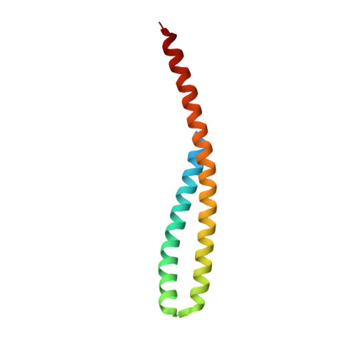
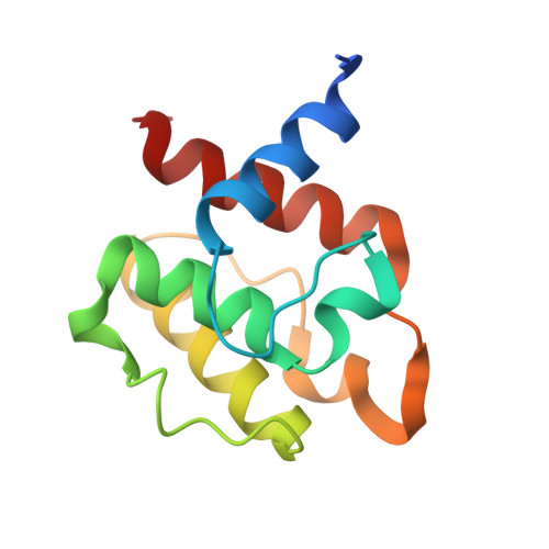
The mechanism of activation of the actin binding protein EHBP1 by Rab8 family members.

Rai, A.Bleimling, N.Vetter, I.R.Goody, R.S.

(2020) Nat Commun 11: 4187-4187

  • DOI: https://doi.org/10.1038/s41467-020-17792-3
  • Primary Citation of Related Structures:  
    6ZSH, 6ZSI, 6ZSJ

  • PubMed Abstract: 

    EHBP1 is an adaptor protein that regulates vesicular trafficking by recruiting Rab8 family members and Eps15-homology domain-containing proteins 1/2 (EHD1/2). It also links endosomes to the actin cytoskeleton. However, the underlying molecular mechanism of activation of EHBP1 actin-binding activity is unclear. Here, we show that both termini of EHBP1 have membrane targeting potential. EHBP1 associates with PI(3)P, PI(5)P, and phosphatidylserine via its N-terminal C2 domain. We show that in the absence of Rab8 family members, the C-terminal bivalent Mical/EHBP Rab binding (bMERB) domain forms an intramolecular complex with its central calponin homology (CH) domain and auto-inhibits actin binding. Rab8 binding to the bMERB domain relieves this inhibition. We have analyzed the CH:bMERB auto-inhibited complex and the active bMERB:Rab8 complex biochemically and structurally. Together with structure-based mutational studies, this explains how binding of Rab8 frees the CH domain and allows it to interact with the actin cytoskeleton, leading to membrane tubulation.


  • Organizational Affiliation

    Department of Structural Biochemistry, Max Planck Institute of Molecular Physiology, Otto-Hahn-Strasse 11, 44227, Dortmund, Germany. amrita.rai@mpi-dortmund.mpg.de.


Macromolecules
Find similar proteins by:  (by identity cutoff)  |  3D Structure
Entity ID: 1
MoleculeChains Sequence LengthOrganismDetailsImage
EH domain-binding protein 1
A, C
106Homo sapiensMutation(s): 0 
Gene Names: EHBP1KIAA0903NACSIN
UniProt & NIH Common Fund Data Resources
Find proteins for Q8NDI1 (Homo sapiens)
Explore Q8NDI1 
Go to UniProtKB:  Q8NDI1
PHAROS:  Q8NDI1
GTEx:  ENSG00000115504 
Entity Groups  
Sequence Clusters30% Identity50% Identity70% Identity90% Identity95% Identity100% Identity
UniProt GroupQ8NDI1
Sequence Annotations
Expand
  • Reference Sequence
Find similar proteins by:  (by identity cutoff)  |  3D Structure
Entity ID: 2
MoleculeChains Sequence LengthOrganismDetailsImage
EH domain-binding protein 1
B, D
114Homo sapiensMutation(s): 0 
Gene Names: EHBP1KIAA0903NACSIN
UniProt & NIH Common Fund Data Resources
Find proteins for Q8NDI1 (Homo sapiens)
Explore Q8NDI1 
Go to UniProtKB:  Q8NDI1
PHAROS:  Q8NDI1
GTEx:  ENSG00000115504 
Entity Groups  
Sequence Clusters30% Identity50% Identity70% Identity90% Identity95% Identity100% Identity
UniProt GroupQ8NDI1
Sequence Annotations
Expand
  • Reference Sequence
Experimental Data & Validation

Experimental Data

  • Method: X-RAY DIFFRACTION
  • Resolution: 2.20 Å
  • R-Value Free: 0.230 
  • R-Value Work: 0.177 
  • R-Value Observed: 0.180 
  • Space Group: P 1 21 1
Unit Cell:
Length ( Å )Angle ( ˚ )
a = 54.06α = 90
b = 48.19β = 97.48
c = 100.3γ = 90
Software Package:
Software NamePurpose
REFMACrefinement
XSCALEdata scaling
PDB_EXTRACTdata extraction
XDSdata reduction
PHASERphasing

Structure Validation

View Full Validation Report


Entry History & Funding Information

Deposition Data


Funding OrganizationLocationGrant Number
Max Planck SocietyGermany--
German Research Foundation (DFG)Germanygrant GO 284/10-1

Revision History  (Full details and data files)

  • Version 1.0: 2020-09-02
    Type: Initial release
  • Version 1.1: 2020-10-14
    Changes: Data collection
  • Version 1.2: 2024-01-31
    Changes: Data collection, Database references, Refinement description