6ZRW

Crystal structure of the fungal lectin CML1


Experimental Data Snapshot

  • Method: X-RAY DIFFRACTION
  • Resolution: 1.35 Å
  • R-Value Free: 0.176 
  • R-Value Work: 0.146 
  • R-Value Observed: 0.147 

wwPDB Validation   3D Report Full Report


This is version 1.3 of the entry. See complete history


Literature

Structure-function relationship of a novel fucoside-binding fruiting body lectin from Coprinopsis cinerea exhibiting nematotoxic activity.

Bleuler-Martinez, S.Varrot, A.Olieric, V.Schubert, M.Vogt, E.Fetz, C.Wohlschlager, T.Plaza, D.F.Walti, M.Duport, Y.Capitani, G.Aebi, M.Kunzler, M.

(2022) Glycobiology 32: 600-615

  • DOI: https://doi.org/10.1093/glycob/cwac020
  • Primary Citation of Related Structures:  
    6ZRW, 6ZU2, 6ZV5

  • PubMed Abstract: 

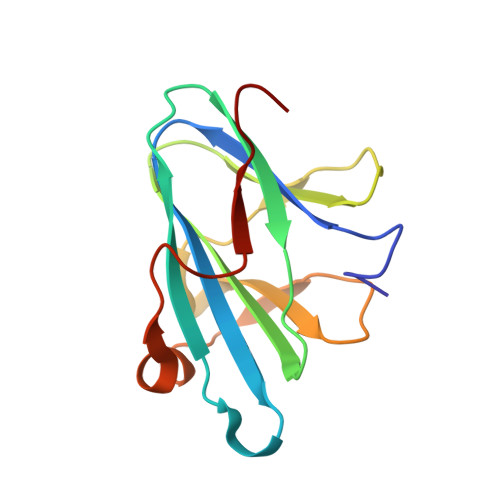
    Lectins are non-immunoglobulin-type proteins that bind to specific carbohydrate epitopes and play important roles in intra- and inter-organismic interactions. Here, we describe a novel fucose-specific lectin, termed CML1, which we identified from fruiting body extracts of Coprinopsis cinerea. For further characterization, the coding sequence for CML1 was cloned and heterologously expressed in Escherichia coli. Feeding of CML1-producing bacteria inhibited larval development of the bacterivorous nematode Caenorhabditis tropicalis, but not of C. elegans. The crystal structure of the recombinant protein in its apo-form and in complex with H type I or Lewis A blood group antigens was determined by X-ray crystallography. The protein folds as a sandwich of 2 antiparallel β-sheets and forms hexamers resulting from a trimer of dimers. The hexameric arrangement was confirmed by small-angle X-ray scattering (SAXS). One carbohydrate-binding site per protomer was found at the dimer interface with both protomers contributing to ligand binding, resulting in a hexavalent lectin. In terms of lectin activity of recombinant CML1, substitution of the carbohydrate-interacting residues His54, Asn55, Trp94, and Arg114 by Ala abolished carbohydrate-binding and nematotoxicity. Although no similarities to any characterized lectin were found, sequence alignments identified many non-characterized agaricomycete proteins. These results suggest that CML1 is the founding member of a novel family of fucoside-binding lectins involved in the defense of agaricomycete fruiting bodies against predation by fungivorous nematodes.


  • Organizational Affiliation

    Institute of Microbiology, Department of Biology, Eidgenössische Technische Hochschule (ETH) Zürich, 8093, Zürich, Switzerland.


Macromolecules
Find similar proteins by:  (by identity cutoff)  |  3D Structure
Entity ID: 1
MoleculeChains Sequence LengthOrganismDetailsImage
Mucin-binding lectin 1
A, B, C, D, E
A, B, C, D, E, F
126Coprinopsis cinereaMutation(s): 0 
Gene Names: cml1
UniProt
Find proteins for B3VS76 (Coprinopsis cinerea)
Explore B3VS76 
Go to UniProtKB:  B3VS76
Entity Groups  
Sequence Clusters30% Identity50% Identity70% Identity90% Identity95% Identity100% Identity
UniProt GroupB3VS76
Sequence Annotations
Expand
  • Reference Sequence
Small Molecules
Ligands 5 Unique
IDChains Name / Formula / InChI Key2D Diagram3D Interactions
PC4
Query on PC4

Download Ideal Coordinates CCD File 
DA [auth D],
EA [auth D],
Q [auth B],
UA [auth F]
TETRACHLOROPLATINATE(II)
Cl4 Pt
SVZRVTAEYVVVPM-UHFFFAOYSA-J
P3C
Query on P3C

Download Ideal Coordinates CCD File 
J [auth A],
K [auth A],
NA [auth E],
V [auth C]
TRICHLOROPLATINATE
Cl3 Pt
UGPIIBDOUTZDQE-UHFFFAOYSA-K
GOL
Query on GOL

Download Ideal Coordinates CCD File 
AA [auth D]
BA [auth D]
CA [auth D]
G [auth A]
H [auth A]
AA [auth D],
BA [auth D],
CA [auth D],
G [auth A],
H [auth A],
I [auth A],
KA [auth E],
LA [auth E],
MA [auth E],
O [auth B],
P [auth B],
S [auth C],
SA [auth F],
T [auth C],
TA [auth F],
U [auth C]
GLYCEROL
C3 H8 O3
PEDCQBHIVMGVHV-UHFFFAOYSA-N
ACT
Query on ACT

Download Ideal Coordinates CCD File 
GA [auth D]
HA [auth D]
IA [auth D]
JA [auth D]
M [auth A]
GA [auth D],
HA [auth D],
IA [auth D],
JA [auth D],
M [auth A],
N [auth A],
QA [auth E],
RA [auth E],
X [auth C],
XA [auth F],
Y [auth C],
YA [auth F],
Z [auth C]
ACETATE ION
C2 H3 O2
QTBSBXVTEAMEQO-UHFFFAOYSA-M
K
Query on K

Download Ideal Coordinates CCD File 
FA [auth D]
L [auth A]
OA [auth E]
PA [auth E]
R [auth B]
FA [auth D],
L [auth A],
OA [auth E],
PA [auth E],
R [auth B],
VA [auth F],
W [auth C],
WA [auth F]
POTASSIUM ION
K
NPYPAHLBTDXSSS-UHFFFAOYSA-N
Experimental Data & Validation

Experimental Data

Unit Cell:
Length ( Å )Angle ( ˚ )
a = 44.497α = 90
b = 73.626β = 101.03
c = 111.956γ = 90
Software Package:
Software NamePurpose
XDSdata scaling
SHELXDEphasing
PHENIXrefinement
Cootmodel building

Structure Validation

View Full Validation Report


Entry History & Funding Information

Deposition Data


Funding OrganizationLocationGrant Number
Swiss National Science FoundationSwitzerland31003A_173097

Revision History  (Full details and data files)

  • Version 1.0: 2021-07-28
    Type: Initial release
  • Version 1.1: 2022-04-06
    Changes: Advisory, Database references
  • Version 1.2: 2022-06-22
    Changes: Database references
  • Version 1.3: 2023-03-01
    Changes: Advisory