6ZPQ

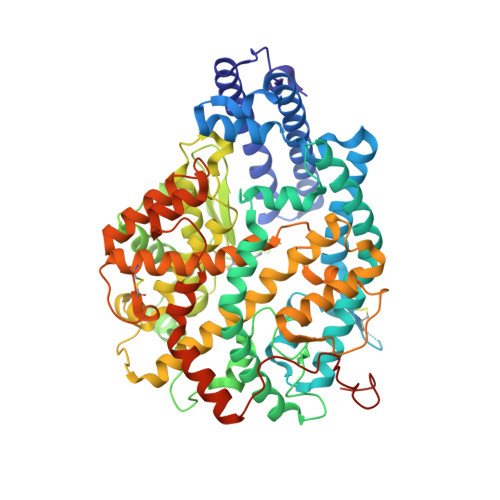
Crystal structure of the open conformation of Angiotensin-1 converting enzyme N-domain.


Experimental Data Snapshot

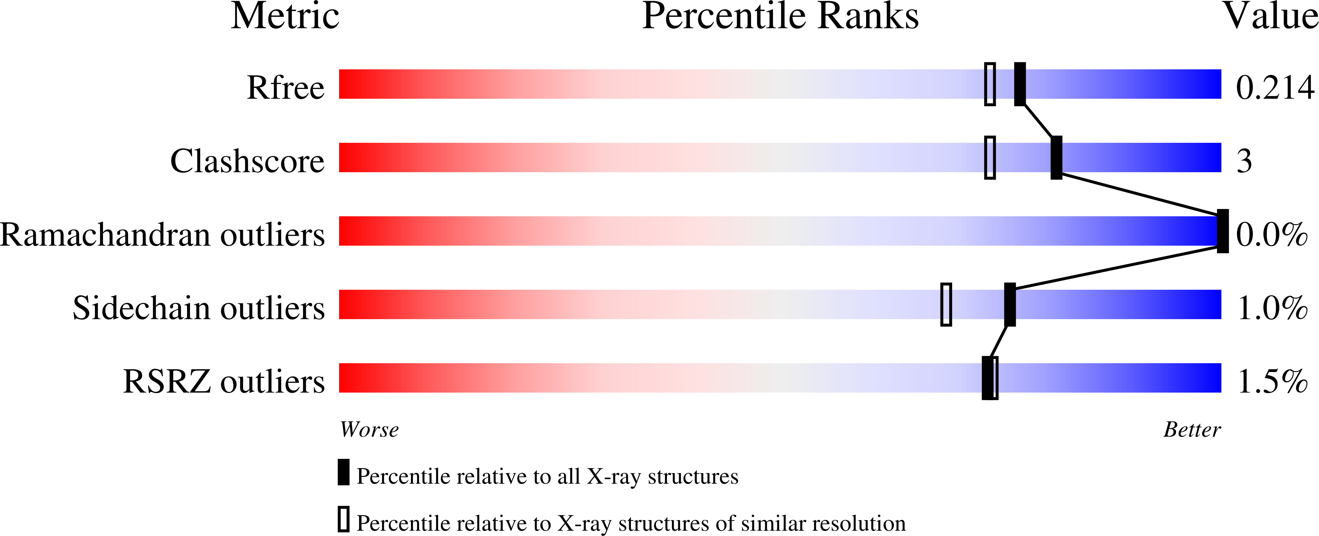
  • Method: X-RAY DIFFRACTION
  • Resolution: 1.85 Å
  • R-Value Free: 0.214 
  • R-Value Work: 0.184 
  • R-Value Observed: 0.185 

wwPDB Validation   3D Report Full Report


Ligand Structure Quality Assessment 


This is version 1.2 of the entry. See complete history


Literature

Angiotensin-converting enzyme open for business: structural insights into the subdomain dynamics.

Cozier, G.E.Lubbe, L.Sturrock, E.D.Acharya, K.R.

(2021) FEBS J 288: 2238-2256

  • DOI: https://doi.org/10.1111/febs.15601
  • Primary Citation of Related Structures:  
    6ZPQ, 6ZPT, 6ZPU

  • PubMed Abstract: 

    Angiotensin-1-converting enzyme (ACE) is a key enzyme in the renin-angiotensin-aldosterone and kinin systems where it cleaves angiotensin I and bradykinin peptides, respectively. However, ACE also participates in numerous other physiological functions, can hydrolyse many peptide substrates and has various exo- and endopeptidase activities. ACE achieves this complexity by containing two homologous catalytic domains (N- and C-domains), which exhibit different substrate specificities. Here, we present the first open conformation structures of ACE N-domain and a unique closed C-domain structure (2.0 Å) where the C terminus of a symmetry-related molecule is observed inserted into the active-site cavity and binding to the zinc ion. The open native N-domain structure (1.85 Å) enables comparison with ACE2, a homologue previously observed in open and closed states. An open S 2 _S'-mutant N-domain structure (2.80 Å) includes mutated residues in the S 2 and S' subsites that effect ligand binding, but are distal to the binding site. Analysis of these structures provides important insights into how structural features of the ACE domains are able to accommodate the wide variety of substrates and allow different peptidase activities. DATABASE: The atomic coordinates and structure factors for Open nACE, Open S2_S'-nACE and Native G13-cACE structures have been deposited with codes 6ZPQ, 6ZPT and 6ZPU, respectively, in the RCSB Protein Data Bank, www.pdb.org.


  • Organizational Affiliation

    Department of Biology and Biochemistry, University of Bath, Bath, UK.


Macromolecules
Find similar proteins by:  (by identity cutoff)  |  3D Structure
Entity ID: 1
MoleculeChains Sequence LengthOrganismDetailsImage
Angiotensin-converting enzyme
A, B, C, D
629Homo sapiensMutation(s): 8 
Gene Names: ACEDCPDCP1
EC: 3.2.1 (PDB Primary Data), 3.4.15.1 (PDB Primary Data)
UniProt & NIH Common Fund Data Resources
Find proteins for P12821 (Homo sapiens)
Explore P12821 
Go to UniProtKB:  P12821
PHAROS:  P12821
GTEx:  ENSG00000159640 
Entity Groups  
Sequence Clusters30% Identity50% Identity70% Identity90% Identity95% Identity100% Identity
UniProt GroupP12821
Sequence Annotations
Expand
  • Reference Sequence
Oligosaccharides

Help

Entity ID: 2
MoleculeChains Length2D Diagram Glycosylation3D Interactions
2-acetamido-2-deoxy-beta-D-glucopyranose-(1-4)-2-acetamido-2-deoxy-beta-D-glucopyranose
E, H, J
2N-Glycosylation
Glycosylation Resources
GlyTouCan:  G42666HT
GlyCosmos:  G42666HT
GlyGen:  G42666HT
Entity ID: 3
MoleculeChains Length2D Diagram Glycosylation3D Interactions
2-acetamido-2-deoxy-beta-D-glucopyranose-(1-4)-[alpha-L-fucopyranose-(1-6)]2-acetamido-2-deoxy-beta-D-glucopyranose
F, G, I, K
3N-Glycosylation
Glycosylation Resources
GlyTouCan:  G21290RB
GlyCosmos:  G21290RB
GlyGen:  G21290RB
Small Molecules
Ligands 12 Unique
IDChains Name / Formula / InChI Key2D Diagram3D Interactions
NAG
Query on NAG

Download Ideal Coordinates CCD File 
CA [auth B]2-acetamido-2-deoxy-beta-D-glucopyranose
C8 H15 N O6
OVRNDRQMDRJTHS-FMDGEEDCSA-N
BCN
Query on BCN

Download Ideal Coordinates CCD File 
DB [auth D],
EA [auth B],
M [auth A],
O [auth A],
SA [auth C]
BICINE
C6 H13 N O4
FSVCELGFZIQNCK-UHFFFAOYSA-N
PGE
Query on PGE

Download Ideal Coordinates CCD File 
EB [auth D],
GA [auth B],
HA [auth B],
R [auth A],
V [auth A]
TRIETHYLENE GLYCOL
C6 H14 O4
ZIBGPFATKBEMQZ-UHFFFAOYSA-N
PEG
Query on PEG

Download Ideal Coordinates CCD File 
FA [auth B],
LA [auth B],
MA [auth B],
P [auth A]
DI(HYDROXYETHYL)ETHER
C4 H10 O3
MTHSVFCYNBDYFN-UHFFFAOYSA-N
MXE
Query on MXE

Download Ideal Coordinates CCD File 
KA [auth B],
TA [auth C]
2-METHOXYETHANOL
C3 H8 O2
XNWFRZJHXBZDAG-UHFFFAOYSA-N
ZN
Query on ZN

Download Ideal Coordinates CCD File 
LB [auth D],
NA [auth B],
Y [auth A],
YA [auth C]
ZINC ION
Zn
PTFCDOFLOPIGGS-UHFFFAOYSA-N
EDO
Query on EDO

Download Ideal Coordinates CCD File 
FB [auth D]
GB [auth D]
HB [auth D]
IA [auth B]
IB [auth D]
FB [auth D],
GB [auth D],
HB [auth D],
IA [auth B],
IB [auth D],
JA [auth B],
Q [auth A],
T [auth A],
U [auth A],
VA [auth C],
W [auth A],
WA [auth C],
X [auth A],
XA [auth C]
1,2-ETHANEDIOL
C2 H6 O2
LYCAIKOWRPUZTN-UHFFFAOYSA-N
BO3
Query on BO3

Download Ideal Coordinates CCD File 
CB [auth D]
DA [auth B]
JB [auth D]
KB [auth D]
L [auth A]
CB [auth D],
DA [auth B],
JB [auth D],
KB [auth D],
L [auth A],
N [auth A],
S [auth A],
UA [auth C]
BORIC ACID
B H3 O3
KGBXLFKZBHKPEV-UHFFFAOYSA-N
CA
Query on CA

Download Ideal Coordinates CCD File 
BA [auth A],
BB [auth C],
NB [auth D],
QA [auth B]
CALCIUM ION
Ca
BHPQYMZQTOCNFJ-UHFFFAOYSA-N
CL
Query on CL

Download Ideal Coordinates CCD File 
MB [auth D],
OA [auth B],
Z [auth A],
ZA [auth C]
CHLORIDE ION
Cl
VEXZGXHMUGYJMC-UHFFFAOYSA-M
MG
Query on MG

Download Ideal Coordinates CCD File 
AA [auth A],
AB [auth C],
PA [auth B]
MAGNESIUM ION
Mg
JLVVSXFLKOJNIY-UHFFFAOYSA-N
NA
Query on NA

Download Ideal Coordinates CCD File 
RA [auth B]SODIUM ION
Na
FKNQFGJONOIPTF-UHFFFAOYSA-N
Experimental Data & Validation

Experimental Data

  • Method: X-RAY DIFFRACTION
  • Resolution: 1.85 Å
  • R-Value Free: 0.214 
  • R-Value Work: 0.184 
  • R-Value Observed: 0.185 
  • Space Group: P 1
Unit Cell:
Length ( Å )Angle ( ˚ )
a = 74.683α = 97.756
b = 99.934β = 90.206
c = 128.701γ = 111.057
Software Package:
Software NamePurpose
PHENIXrefinement
PHENIXrefinement
DIALSdata reduction
Aimlessdata scaling
PHASERphasing

Structure Validation

View Full Validation Report


Ligand Structure Quality Assessment 


Entry History & Funding Information

Deposition Data


Funding OrganizationLocationGrant Number
Medical Research Council (MRC, United Kingdom)United KingdomMR/M026647/1

Revision History  (Full details and data files)

  • Version 1.0: 2020-10-28
    Type: Initial release
  • Version 1.1: 2021-04-14
    Changes: Database references
  • Version 1.2: 2024-01-31
    Changes: Data collection, Database references, Refinement description