6ZPE

Nonstructural protein 10 (nsp10) from SARS CoV-2


Experimental Data Snapshot

  • Method: X-RAY DIFFRACTION
  • Resolution: 1.58 Å
  • R-Value Free: 0.160 
  • R-Value Work: 0.151 
  • R-Value Observed: 0.152 

wwPDB Validation   3D Report Full Report


This is version 1.2 of the entry. See complete history


Literature

Crystal Structure of Non-Structural Protein 10 from Severe Acute Respiratory Syndrome Coronavirus-2.

Rogstam, A.Nyblom, M.Christensen, S.Sele, C.Talibov, V.O.Lindvall, T.Rasmussen, A.A.Andre, I.Fisher, Z.Knecht, W.Kozielski, F.

(2020) Int J Mol Sci 21

  • DOI: https://doi.org/10.3390/ijms21197375
  • Primary Citation of Related Structures:  
    6ZCT, 6ZPE

  • PubMed Abstract: 

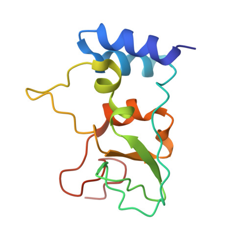
    Severe Acute Respiratory Syndrome Coronavirus-2 (SARS-CoV-2), causing Coronavirus Disease 19 (COVID-19), emerged at the end of 2019 and quickly spread to cause a global pandemic with severe socio-economic consequences. The early sequencing of its RNA genome revealed its high similarity to SARS, likely to have originated from bats. The SARS-CoV-2 non-structural protein 10 (nsp10) displays high sequence similarity with its SARS homologue, which binds to and stimulates the 3'-to-5' exoribonuclease and the 2'-O-methlytransferase activities of nsps 14 and 16, respectively. Here, we report the biophysical characterization and 1.6 Å resolution structure of the unbound form of nsp10 from SARS-CoV-2 and compare it to the structures of its SARS homologue and the complex-bound form with nsp16 from SARS-CoV-2. The crystal structure and solution behaviour of nsp10 will not only form the basis for understanding the role of SARS-CoV-2 nsp10 as a central player of the viral RNA capping apparatus, but will also serve as a basis for the development of inhibitors of nsp10, interfering with crucial functions of the replication-transcription complex and virus replication.


  • Organizational Affiliation

    Department of Biology & Lund Protein Production Platform, Lund University, Sölvegatan 35, 22362 Lund, Sweden.


Macromolecules
Find similar proteins by:  (by identity cutoff)  |  3D Structure
Entity ID: 1
MoleculeChains Sequence LengthOrganismDetailsImage
Replicase polyprotein 1ab125Severe acute respiratory syndrome coronavirus 2Mutation(s): 0 
Gene Names: rep1a-1b
EC: 3.4.19.12 (PDB Primary Data), 3.4.22 (PDB Primary Data), 3.4.22.69 (PDB Primary Data), 2.7.7.48 (PDB Primary Data), 3.6.4.12 (PDB Primary Data), 3.6.4.13 (PDB Primary Data), 3.1.13 (PDB Primary Data), 3.1 (PDB Primary Data), 2.1.1 (PDB Primary Data)
UniProt
Find proteins for P0DTD1 (Severe acute respiratory syndrome coronavirus 2)
Explore P0DTD1 
Go to UniProtKB:  P0DTD1
Entity Groups  
Sequence Clusters30% Identity50% Identity70% Identity90% Identity95% Identity100% Identity
UniProt GroupP0DTD1
Sequence Annotations
Expand
  • Reference Sequence
Experimental Data & Validation

Experimental Data

  • Method: X-RAY DIFFRACTION
  • Resolution: 1.58 Å
  • R-Value Free: 0.160 
  • R-Value Work: 0.151 
  • R-Value Observed: 0.152 
  • Space Group: I 21 3
Unit Cell:
Length ( Å )Angle ( ˚ )
a = 106.624α = 90
b = 106.624β = 90
c = 106.624γ = 90
Software Package:
Software NamePurpose
XDSdata reduction
Aimlessdata scaling
PHENIXrefinement
PDB_EXTRACTdata extraction
PHASERphasing

Structure Validation

View Full Validation Report


Entry History 

Deposition Data

Revision History  (Full details and data files)

  • Version 1.0: 2020-10-14
    Type: Initial release
  • Version 1.1: 2020-10-21
    Changes: Database references
  • Version 1.2: 2024-01-31
    Changes: Data collection, Database references, Refinement description