6Z8L

Alpha-Amylase in complex with probe fragments


Experimental Data Snapshot

  • Method: X-RAY DIFFRACTION
  • Resolution: 1.40 Å
  • R-Value Free: 0.154 
  • R-Value Work: 0.142 
  • R-Value Observed: 0.143 

wwPDB Validation   3D Report Full Report


Ligand Structure Quality Assessment 


This is version 1.2 of the entry. See complete history


Literature

Enhancing glycan stability via site-selective fluorination: modulating substrate orientation by molecular design.

Axer, A.Jumde, R.P.Adam, S.Faust, A.Schafers, M.Fobker, M.Koehnke, J.Hirsch, A.K.H.Gilmour, R.

(2020) Chem Sci 12: 1286-1294

  • DOI: https://doi.org/10.1039/d0sc04297h
  • Primary Citation of Related Structures:  
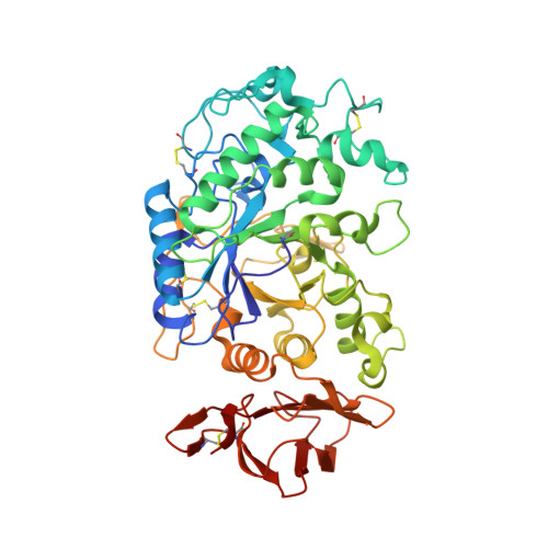
    6Z8L

  • PubMed Abstract: 

    Single site OH → F substitution at the termini of maltotetraose leads to significantly improved hydrolytic stability towards α-amylase and α-glucosidase relative to the natural compound. To explore the effect of molecular editing, selectively modified oligosaccharides were prepared via a convergent α-selective strategy. Incubation experiments in purified α-amylase and α-glucosidase, and in human and murine blood serum, provide insight into the influence of fluorine on the hydrolytic stability of these clinically important scaffolds. Enhancements of ca . 1 order of magnitude result from these subtle single point mutations. Modification at the monosaccharide furthest from the probable enzymatic cleavage termini leads to the greatest improvement in stability. In the case of α-amylase, docking studies revealed that retentive C2-fluorination at the reducing end inverts the orientation in which the substrate is bound. A co-crystal structure of human α-amylase revealed maltose units bound at the active-site. In view of the evolving popularity of C(sp 3 )-F bioisosteres in medicinal chemistry, and the importance of maltodextrins in bacterial imaging, this discovery begins to reconcile the information-rich nature of carbohydrates with their intrinsic hydrolytic vulnerabilities.


  • Organizational Affiliation

    Organisch Chemisches Institut, WWU Münster Corrensstraße 36 48149 Münster Germany.


Macromolecules
Find similar proteins by:  (by identity cutoff)  |  3D Structure
Entity ID: 1
MoleculeChains Sequence LengthOrganismDetailsImage
Pancreatic alpha-amylase496Homo sapiensMutation(s): 0 
EC: 3.2.1.1
UniProt & NIH Common Fund Data Resources
Find proteins for P04746 (Homo sapiens)
Explore P04746 
Go to UniProtKB:  P04746
PHAROS:  P04746
GTEx:  ENSG00000243480 
Entity Groups  
Sequence Clusters30% Identity50% Identity70% Identity90% Identity95% Identity100% Identity
UniProt GroupP04746
Sequence Annotations
Expand
  • Reference Sequence
Oligosaccharides

Help

Entity ID: 2
MoleculeChains Length2D Diagram Glycosylation3D Interactions
alpha-D-glucopyranose-(1-4)-alpha-D-glucopyranose
B, C, D, E
2N/A
Glycosylation Resources
GlyTouCan:  G07411ON
GlyCosmos:  G07411ON
Small Molecules
Modified Residues  1 Unique
IDChains TypeFormula2D DiagramParent
PCA
Query on PCA
A
L-PEPTIDE LINKINGC5 H7 N O3GLN
Experimental Data & Validation

Experimental Data

  • Method: X-RAY DIFFRACTION
  • Resolution: 1.40 Å
  • R-Value Free: 0.154 
  • R-Value Work: 0.142 
  • R-Value Observed: 0.143 
  • Space Group: P 21 21 21
Unit Cell:
Length ( Å )Angle ( ˚ )
a = 51.873α = 90
b = 73.729β = 90
c = 135.207γ = 90
Software Package:
Software NamePurpose
MolProbitymodel building
PHENIXrefinement
MolProbitymodel building
XDSdata reduction
SCALAdata scaling
PHASERphasing

Structure Validation

View Full Validation Report


Ligand Structure Quality Assessment 


Entry History & Funding Information

Deposition Data


Funding OrganizationLocationGrant Number
German Research Foundation (DFG)KO 4116/3-1

Revision History  (Full details and data files)

  • Version 1.0: 2020-12-02
    Type: Initial release
  • Version 1.1: 2021-07-07
    Changes: Database references
  • Version 1.2: 2024-01-24
    Changes: Data collection, Database references, Refinement description