


Crystal structure of Paradendryphiella salina PL7A alginate lyase
Wilkens, C., Fredslund, F., Welner, D.H.To be published.
Experimental Data Snapshot
wwPDB Validation 3D Report Full Report

Entity ID: 1 | |||||
|---|---|---|---|---|---|
| Molecule | Chains | Sequence Length | Organism | Details | Image |
| Alginate lyase (PL7) | 231 | Paradendryphiella salina | Mutation(s): 0 Gene Names: PsAlg7A | 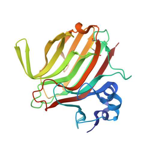 | |
UniProt | |||||
Find proteins for A0A485PVH1 (Paradendryphiella salina) Explore A0A485PVH1 Go to UniProtKB: A0A485PVH1 | |||||
Entity Groups | |||||
| Sequence Clusters | 30% Identity50% Identity70% Identity90% Identity95% Identity100% Identity | ||||
| UniProt Group | A0A485PVH1 | ||||
Sequence AnnotationsExpand | |||||
| |||||
| Length ( Å ) | Angle ( ˚ ) |
|---|---|
| a = 34.768 | α = 90 |
| b = 80.442 | β = 90 |
| c = 81.375 | γ = 90 |
| Software Name | Purpose |
|---|---|
| PHENIX | refinement |
| XDS | data reduction |
| Aimless | data scaling |
| PHASER | phasing |
RCSB PDB (citation) is hosted by
RCSB PDB is a member of the

