6YQH

GH146 beta-L-arabinofuranosidase bound to covalent inhibitor


Experimental Data Snapshot

  • Method: X-RAY DIFFRACTION
  • Resolution: 1.41 Å
  • R-Value Free: 0.170 
  • R-Value Work: 0.129 

wwPDB Validation   3D Report Full Report


Ligand Structure Quality Assessment 


This is version 1.3 of the entry. See complete history


Literature

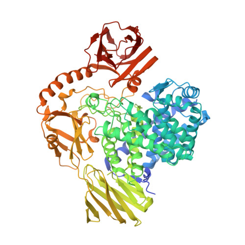
Cysteine Nucleophiles in Glycosidase Catalysis: Application of a Covalent beta-l-Arabinofuranosidase Inhibitor.

McGregor, N.G.S.Coines, J.Borlandelli, V.Amaki, S.Artola, M.Nin-Hill, A.Linzel, D.Yamada, C.Arakawa, T.Ishiwata, A.Ito, Y.van der Marel, G.A.Codee, J.D.C.Fushinobu, S.Overkleeft, H.S.Rovira, C.Davies, G.J.

(2021) Angew Chem Int Ed Engl 60: 5754-5758

  • DOI: https://doi.org/10.1002/anie.202013920
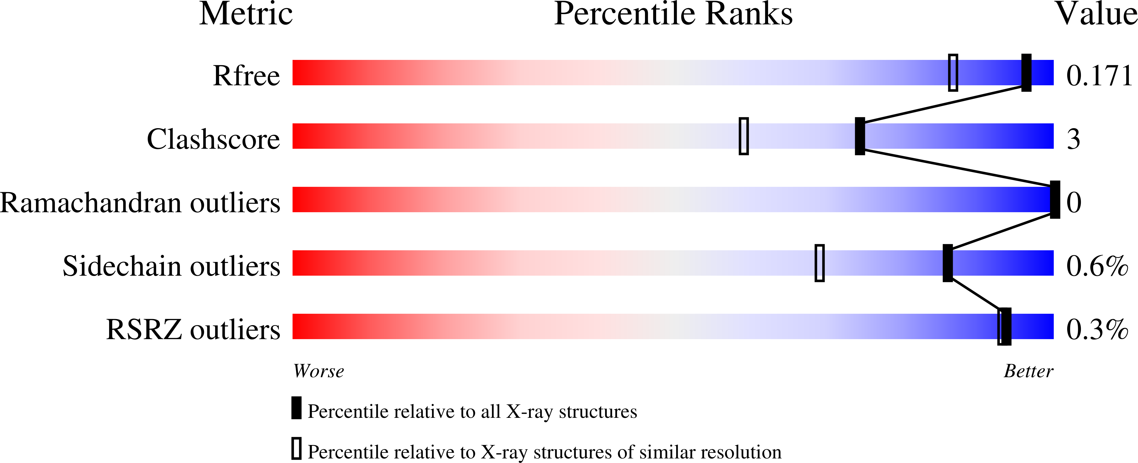
  • Primary Citation of Related Structures:  
    6YQH, 7BZL, 7DIF

  • PubMed Abstract: 

    The recent discovery of zinc-dependent retaining glycoside hydrolases (GHs), with active sites built around a Zn(Cys) 3 (Glu) coordination complex, has presented unresolved mechanistic questions. In particular, the proposed mechanism, depending on a Zn-coordinated cysteine nucleophile and passing through a thioglycosyl enzyme intermediate, remains controversial. This is primarily due to the expected stability of the intermediate C-S bond. To facilitate the study of this atypical mechanism, we report the synthesis of a cyclophellitol-derived β-l-arabinofuranosidase inhibitor, hypothesised to react with the catalytic nucleophile to form a non-hydrolysable adduct analogous to the mechanistic covalent intermediate. This β-l-arabinofuranosidase inhibitor reacts exclusively with the proposed cysteine thiol catalytic nucleophiles of representatives of GH families 127 and 146. X-ray crystal structures determined for the resulting adducts enable MD and QM/MM simulations, which provide insight into the mechanism of thioglycosyl enzyme intermediate breakdown. Leveraging the unique chemistry of cyclophellitol derivatives, the structures and simulations presented here support the assignment of a zinc-coordinated cysteine as the catalytic nucleophile and illuminate the finely tuned energetics of this remarkable metalloenzyme clan.


  • Organizational Affiliation

    York Structural Biology Laboratory, Department of Chemistry, The University of York, Heslington, York, YO10 5DD, UK.


Macromolecules
Find similar proteins by:  (by identity cutoff)  |  3D Structure
Entity ID: 1
MoleculeChains Sequence LengthOrganismDetailsImage
Acetyl-CoA carboxylase, biotin carboxylaseA [auth AAA]802Bacteroides thetaiotaomicron VPI-5482Mutation(s): 0 
Gene Names: BT_0349
UniProt
Find proteins for Q8AAW3 (Bacteroides thetaiotaomicron (strain ATCC 29148 / DSM 2079 / JCM 5827 / CCUG 10774 / NCTC 10582 / VPI-5482 / E50))
Explore Q8AAW3 
Go to UniProtKB:  Q8AAW3
Entity Groups  
Sequence Clusters30% Identity50% Identity70% Identity90% Identity95% Identity100% Identity
UniProt GroupQ8AAW3
Sequence Annotations
Expand
  • Reference Sequence
Small Molecules
Ligands 6 Unique
IDChains Name / Formula / InChI Key2D Diagram3D Interactions
MES
Query on MES

Download Ideal Coordinates CCD File 
F [auth AAA]2-(N-MORPHOLINO)-ETHANESULFONIC ACID
C6 H13 N O4 S
SXGZJKUKBWWHRA-UHFFFAOYSA-N
PGE
Query on PGE

Download Ideal Coordinates CCD File 
D [auth AAA]TRIETHYLENE GLYCOL
C6 H14 O4
ZIBGPFATKBEMQZ-UHFFFAOYSA-N
PJ5 (Subject of Investigation/LOI)
Query on PJ5

Download Ideal Coordinates CCD File 
E [auth AAA](1~{S},2~{S},3~{S},4~{S})-4-(hydroxymethyl)cyclopentane-1,2,3-triol
C6 H12 O4
PHKHGSSZAJVEQK-BXKVDMCESA-N
PEG
Query on PEG

Download Ideal Coordinates CCD File 
B [auth AAA],
C [auth AAA]
DI(HYDROXYETHYL)ETHER
C4 H10 O3
MTHSVFCYNBDYFN-UHFFFAOYSA-N
ZN
Query on ZN

Download Ideal Coordinates CCD File 
U [auth AAA]ZINC ION
Zn
PTFCDOFLOPIGGS-UHFFFAOYSA-N
FMT
Query on FMT

Download Ideal Coordinates CCD File 
G [auth AAA]
H [auth AAA]
I [auth AAA]
J [auth AAA]
K [auth AAA]
G [auth AAA],
H [auth AAA],
I [auth AAA],
J [auth AAA],
K [auth AAA],
L [auth AAA],
M [auth AAA],
N [auth AAA],
O [auth AAA],
P [auth AAA],
Q [auth AAA],
R [auth AAA],
S [auth AAA],
T [auth AAA]
FORMIC ACID
C H2 O2
BDAGIHXWWSANSR-UHFFFAOYSA-N
Experimental Data & Validation

Experimental Data

  • Method: X-RAY DIFFRACTION
  • Resolution: 1.41 Å
  • R-Value Free: 0.170 
  • R-Value Work: 0.129 
  • Space Group: P 43 21 2
Unit Cell:
Length ( Å )Angle ( ˚ )
a = 95.418α = 90
b = 95.418β = 90
c = 189.068γ = 90
Software Package:
Software NamePurpose
REFMACrefinement
xia2data reduction
xia2data scaling
PHASERphasing

Structure Validation

View Full Validation Report


Ligand Structure Quality Assessment 


Entry History & Funding Information

Deposition Data


Funding OrganizationLocationGrant Number
Biotechnology and Biological Sciences Research Council (BBSRC)United KingdomBB/R001162/1

Revision History  (Full details and data files)

  • Version 1.0: 2021-01-27
    Type: Initial release
  • Version 1.1: 2021-02-24
    Changes: Database references
  • Version 1.2: 2021-03-10
    Changes: Database references
  • Version 1.3: 2024-01-24
    Changes: Data collection, Database references, Derived calculations, Refinement description