6YHU

Crystal structure of the nsp7-nsp8 complex of SARS-CoV-2


Experimental Data Snapshot

  • Method: X-RAY DIFFRACTION
  • Resolution: 2.00 Å
  • R-Value Free: 0.239 
  • R-Value Work: 0.214 
  • R-Value Observed: 0.216 

wwPDB Validation   3D Report Full Report


This is version 1.3 of the entry. See complete history


Literature

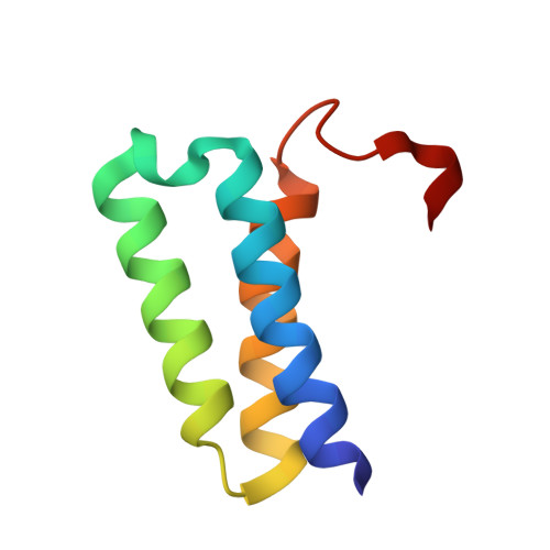
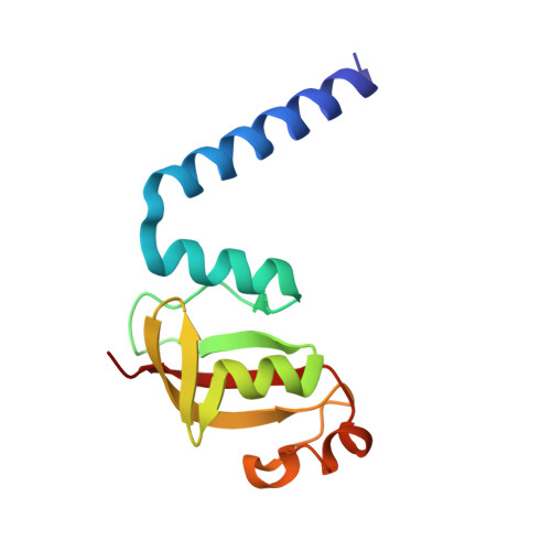
Structural analysis of the putative SARS-CoV-2 primase complex.

Konkolova, E.Klima, M.Nencka, R.Boura, E.

(2020) J Struct Biol 211: 107548-107548

  • DOI: https://doi.org/10.1016/j.jsb.2020.107548
  • Primary Citation of Related Structures:  
    6YHU

  • PubMed Abstract: 

    We report the crystal structure of the SARS-CoV-2 putative primase composed of the nsp7 and nsp8 proteins. We observed a dimer of dimers (2:2 nsp7-nsp8) in the crystallographic asymmetric unit. The structure revealed a fold with a helical core of the heterotetramer formed by both nsp7 and nsp8 that is flanked with two symmetry-related nsp8 β-sheet subdomains. It was also revealed that two hydrophobic interfaces one of approx. 1340 Å 2 connects the nsp7 to nsp8 and a second one of approx. 950 Å 2 connects the dimers and form the observed heterotetramer. Interestingly, analysis of the surface electrostatic potential revealed a putative RNA binding site that is formed only within the heterotetramer.


  • Organizational Affiliation

    Institute of Organic Chemistry and Biochemistry AS CR, v.v.i, Flemingovo nam. 2, 166 10 Prague 6, Czech Republic.


Macromolecules
Find similar proteins by:  (by identity cutoff)  |  3D Structure
Entity ID: 1
MoleculeChains Sequence LengthOrganismDetailsImage
Replicase polyprotein 1a
A, C
71Severe acute respiratory syndrome coronavirus 2Mutation(s): 0 
EC: 3.4.19.12 (PDB Primary Data), 3.4.22 (PDB Primary Data), 3.4.22.69 (PDB Primary Data)
UniProt
Find proteins for P0DTD1 (Severe acute respiratory syndrome coronavirus 2)
Explore P0DTD1 
Go to UniProtKB:  P0DTD1
Entity Groups  
Sequence Clusters30% Identity50% Identity70% Identity90% Identity95% Identity100% Identity
UniProt GroupP0DTD1
Sequence Annotations
Expand
  • Reference Sequence
Find similar proteins by:  (by identity cutoff)  |  3D Structure
Entity ID: 2
MoleculeChains Sequence LengthOrganismDetailsImage
Replicase polyprotein 1a
B, D
117Severe acute respiratory syndrome coronavirus 2Mutation(s): 0 
EC: 3.4.19.12 (PDB Primary Data), 3.4.22 (PDB Primary Data), 3.4.22.69 (PDB Primary Data)
UniProt
Find proteins for P0DTD1 (Severe acute respiratory syndrome coronavirus 2)
Explore P0DTD1 
Go to UniProtKB:  P0DTD1
Entity Groups  
Sequence Clusters30% Identity50% Identity70% Identity90% Identity95% Identity100% Identity
UniProt GroupP0DTD1
Sequence Annotations
Expand
  • Reference Sequence
Experimental Data & Validation

Experimental Data

  • Method: X-RAY DIFFRACTION
  • Resolution: 2.00 Å
  • R-Value Free: 0.239 
  • R-Value Work: 0.214 
  • R-Value Observed: 0.216 
  • Space Group: P 1 21 1
Unit Cell:
Length ( Å )Angle ( ˚ )
a = 56.42α = 90
b = 49.66β = 106.413
c = 64.75γ = 90
Software Package:
Software NamePurpose
PHENIXrefinement
XDSdata reduction
XDSdata scaling
PHASERphasing

Structure Validation

View Full Validation Report


Entry History 

Deposition Data

Revision History  (Full details and data files)

  • Version 1.0: 2020-04-29
    Type: Initial release
  • Version 1.1: 2020-05-06
    Changes: Database references, Source and taxonomy, Structure summary
  • Version 1.2: 2020-11-11
    Changes: Database references
  • Version 1.3: 2024-01-24
    Changes: Data collection, Database references, Refinement description