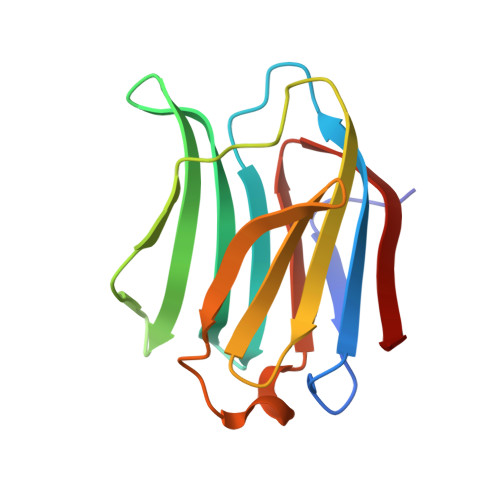
6Y4C

Structure of galectin-3C in complex with lactose determined by serial crystallography using an XtalTool support


Experimental Data Snapshot

  • Method: X-RAY DIFFRACTION
  • Resolution: 1.70 Å
  • R-Value Free: 0.181 
  • R-Value Work: 0.145 
  • R-Value Observed: 0.148 

wwPDB Validation   3D Report Full Report


This is version 2.2 of the entry. See complete history


Literature

Current status and future opportunities for serial crystallography at MAX IV Laboratory.

Shilova, A.Lebrette, H.Aurelius, O.Nan, J.Welin, M.Kovacic, R.Ghosh, S.Safari, C.Friel, R.J.Milas, M.Matej, Z.Hogbom, M.Branden, G.Kloos, M.Shoeman, R.L.Doak, B.Ursby, T.Hakansson, M.Logan, D.T.Mueller, U.

(2020) J Synchrotron Radiat 27: 1095-1102

  • DOI: https://doi.org/10.1107/S1600577520008735
  • Primary Citation of Related Structures:  
    6Y2N, 6Y4C, 6Y78

  • PubMed Abstract: 

    Over the last decade, serial crystallography, a method to collect complete diffraction datasets from a large number of microcrystals delivered and exposed to an X-ray beam in random orientations at room temperature, has been successfully implemented at X-ray free-electron lasers and synchrotron radiation facility beamlines. This development relies on a growing variety of sample presentation methods, including different fixed target supports, injection methods using gas-dynamic virtual-nozzle injectors and high-viscosity extrusion injectors, and acoustic levitation of droplets, each with unique requirements. In comparison with X-ray free-electron lasers, increased beam time availability makes synchrotron facilities very attractive to perform serial synchrotron X-ray crystallography (SSX) experiments. Within this work, the possibilities to perform SSX at BioMAX, the first macromolecular crystallography beamline at  MAX IV Laboratory in Lund, Sweden, are described, together with case studies from the SSX user program: an implementation of a high-viscosity extrusion injector to perform room temperature serial crystallography at BioMAX using two solid supports - silicon nitride membranes (Silson, UK) and XtalTool (Jena Bioscience, Germany). Future perspectives for the dedicated serial crystallography beamline MicroMAX at MAX IV Laboratory, which will provide parallel and intense micrometre-sized X-ray beams, are discussed.


  • Organizational Affiliation

    MAX IV Laboratory, Lund University, Fotongatan 2, Lund 22484, Sweden.


Macromolecules
Find similar proteins by:  (by identity cutoff)  |  3D Structure
Entity ID: 1
MoleculeChains Sequence LengthOrganismDetailsImage
Galectin-3138Homo sapiensMutation(s): 0 
Gene Names: LGALS3MAC2
UniProt & NIH Common Fund Data Resources
Find proteins for P17931 (Homo sapiens)
Explore P17931 
Go to UniProtKB:  P17931
PHAROS:  P17931
GTEx:  ENSG00000131981 
Entity Groups  
Sequence Clusters30% Identity50% Identity70% Identity90% Identity95% Identity100% Identity
UniProt GroupP17931
Sequence Annotations
Expand
  • Reference Sequence
Oligosaccharides

Help

Entity ID: 2
MoleculeChains Length2D Diagram Glycosylation3D Interactions
beta-D-galactopyranose-(1-4)-beta-D-glucopyranose
B
2N/A
Glycosylation Resources
GlyTouCan:  G84224TW
GlyCosmos:  G84224TW
GlyGen:  G84224TW
Small Molecules
Ligands 1 Unique
IDChains Name / Formula / InChI Key2D Diagram3D Interactions
CL
Query on CL

Download Ideal Coordinates CCD File 
C [auth A]CHLORIDE ION
Cl
VEXZGXHMUGYJMC-UHFFFAOYSA-M
Biologically Interesting Molecules (External Reference) 1 Unique
Experimental Data & Validation

Experimental Data

  • Method: X-RAY DIFFRACTION
  • Resolution: 1.70 Å
  • R-Value Free: 0.181 
  • R-Value Work: 0.145 
  • R-Value Observed: 0.148 
  • Space Group: P 21 21 21
Unit Cell:
Length ( Å )Angle ( ˚ )
a = 36.66α = 90
b = 58.05β = 90
c = 63.72γ = 90
Software Package:
Software NamePurpose
PHENIXrefinement
CrystFELdata reduction
CrystFELdata scaling
PHENIXphasing

Structure Validation

View Full Validation Report


Entry History & Funding Information

Deposition Data


Funding OrganizationLocationGrant Number
VinnovaSweden2018-04409

Revision History  (Full details and data files)

  • Version 1.0: 2020-06-17
    Type: Initial release
  • Version 2.0: 2020-07-29
    Type: Remediation
    Reason: Carbohydrate remediation
    Changes: Atomic model, Author supporting evidence, Data collection, Derived calculations, Non-polymer description, Structure summary
  • Version 2.1: 2020-09-16
    Changes: Database references, Structure summary
  • Version 2.2: 2024-01-24
    Changes: Data collection, Database references, Refinement description