Single tryptophan Y160W mutant of homooligomeric E. coli purine nucleoside phosphorylase implies that dimers forming the hexamer are functionally not equivalent.
Narczyk, M., Mioduszewski, L., Oksiejuk, A., Winiewska-Szajewska, M., Wielgus-Kutrowska, B., Gojdz, A., Ciesla, J., Bzowska, A.(2021) Sci Rep 11: 11144-11144
- PubMed: 34045551
- DOI: https://doi.org/10.1038/s41598-021-90472-4
- Primary Citation of Related Structures:
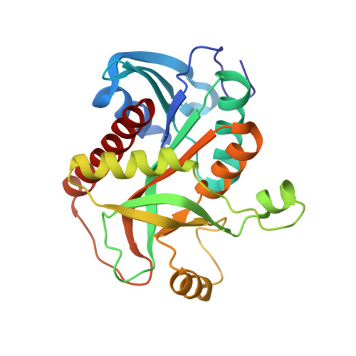
6XZ2 - PubMed Abstract:
E. coli purine nucleoside phosphorylase is a homohexamer, which structure, in the apo form, can be described as a trimer of dimers. Earlier studies suggested that ligand binding and kinetic properties are well described by two binding constants and two sets of kinetic constants. However, most of the crystal structures of this enzyme complexes with ligands do not hold the three-fold symmetry, but only two-fold symmetry, as one of the three dimers is different (both active sites in the open conformation) from the other two (one active site in the open and one in the closed conformation). Our recent detailed studies conducted over broad ligand concentration range suggest that protein-ligand complex formation in solution actually deviates from the two-binding-site model. To reveal the details of interactions present in the hexameric molecule we have engineered a single tryptophan Y160W mutant, responding with substantial intrinsic fluorescence change upon ligand binding. By observing various physical properties of the protein and its various complexes with substrate and substrate analogues we have shown that indeed three-binding-site model is necessary to properly describe binding of ligands by both the wild type enzyme and the Y160W mutant. Thus we have pointed out that a symmetrical dimer with both active sites in the open conformation is not forced to adopt this conformation by interactions in the crystal, but most probably the dimers forming the hexamer in solution are not equivalent as well. This, in turn, implies that an allosteric cooperation occurs not only within a dimer, but also among all three dimers forming a hexameric molecule.
Organizational Affiliation:
Division of Biophysics, Institute of Experimental Physics, Faculty of Physics, University of Warsaw, Pasteura 5, 02-093, Warsaw, Poland.