6XA4

Crystal structure of the SARS-CoV-2 (COVID-19) main protease in complex with UAW241


Experimental Data Snapshot

  • Method: X-RAY DIFFRACTION
  • Resolution: 1.65 Å
  • R-Value Free: 0.239 
  • R-Value Work: 0.198 
  • R-Value Observed: 0.200 

wwPDB Validation   3D Report Full Report


This is version 2.0 of the entry. See complete history


Literature

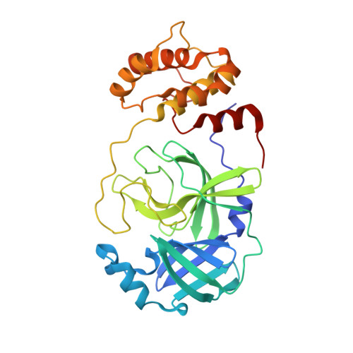
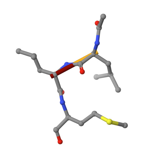
Structure and inhibition of the SARS-CoV-2 main protease reveal strategy for developing dual inhibitors against M pro and cathepsin L.

Sacco, M.D.Ma, C.Lagarias, P.Gao, A.Townsend, J.A.Meng, X.Dube, P.Zhang, X.Hu, Y.Kitamura, N.Hurst, B.Tarbet, B.Marty, M.T.Kolocouris, A.Xiang, Y.Chen, Y.Wang, J.

(2020) Sci Adv 6

  • DOI: https://doi.org/10.1126/sciadv.abe0751
  • Primary Citation of Related Structures:  
    6XA4, 6XBG, 6XBH, 6XBI, 6XFN

  • PubMed Abstract: 

    The main protease (M pro ) of SARS-CoV-2 is a key antiviral drug target. While most M pro inhibitors have a γ-lactam glutamine surrogate at the P1 position, we recently found that several M pro inhibitors have hydrophobic moieties at the P1 site, including calpain inhibitors II and XII, which are also active against human cathepsin L, a host protease that is important for viral entry. In this study, we solved x-ray crystal structures of M pro in complex with calpain inhibitors II and XII and three analogs of GC-376 The structure of M pro with calpain inhibitor II confirmed that the S1 pocket can accommodate a hydrophobic methionine side chain, challenging the idea that a hydrophilic residue is necessary at this position. The structure of calpain inhibitor XII revealed an unexpected, inverted binding pose. Together, the biochemical, computational, structural, and cellular data presented herein provide new directions for the development of dual inhibitors as SARS-CoV-2 antivirals.


  • Organizational Affiliation

    Department of Molecular Medicine, Morsani College of Medicine, University of South Florida, Tampa, FL 33612, USA.


Macromolecules
Find similar proteins by:  (by identity cutoff)  |  3D Structure
Entity ID: 1
MoleculeChains Sequence LengthOrganismDetailsImage
3C-like proteinase308Severe acute respiratory syndrome coronavirus 2Mutation(s): 0 
Gene Names: rep1a-1b
EC: 3.4.22.69
UniProt
Find proteins for P0DTD1 (Severe acute respiratory syndrome coronavirus 2)
Explore P0DTD1 
Go to UniProtKB:  P0DTD1
Entity Groups  
Sequence Clusters30% Identity50% Identity70% Identity90% Identity95% Identity100% Identity
UniProt GroupP0DTD1
Sequence Annotations
Expand
  • Reference Sequence

Find similar proteins by:  Sequence   |   3D Structure  

Entity ID: 2
MoleculeChains Sequence LengthOrganismDetailsImage
inhibitor UAW2414synthetic constructMutation(s): 0 
Sequence Annotations
Expand
  • Reference Sequence
Small Molecules
Ligands 1 Unique
IDChains Name / Formula / InChI Key2D Diagram3D Interactions
GOL
Query on GOL

Download Ideal Coordinates CCD File 
C [auth A]GLYCEROL
C3 H8 O3
PEDCQBHIVMGVHV-UHFFFAOYSA-N
Biologically Interesting Molecules (External Reference) 1 Unique
Experimental Data & Validation

Experimental Data

  • Method: X-RAY DIFFRACTION
  • Resolution: 1.65 Å
  • R-Value Free: 0.239 
  • R-Value Work: 0.198 
  • R-Value Observed: 0.200 
  • Space Group: C 1 2 1
Unit Cell:
Length ( Å )Angle ( ˚ )
a = 113.538α = 90
b = 54.204β = 100.71
c = 45.414γ = 90
Software Package:
Software NamePurpose
REFMACrefinement
HKL-3000data scaling
MOLREPphasing
PDB_EXTRACTdata extraction
HKL-3000data reduction

Structure Validation

View Full Validation Report


Entry History & Funding Information

Deposition Data


Funding OrganizationLocationGrant Number
National Institutes of Health/National Institute Of Allergy and Infectious Diseases (NIH/NIAID)United StatesAI147325

Revision History  (Full details and data files)

  • Version 1.0: 2020-06-17
    Type: Initial release
  • Version 1.1: 2020-11-18
    Changes: Database references
  • Version 1.2: 2020-12-23
    Changes: Database references
  • Version 1.3: 2021-01-27
    Changes: Structure summary
  • Version 1.4: 2023-10-18
    Changes: Data collection, Database references, Refinement description
  • Version 2.0: 2023-11-15
    Changes: Atomic model, Data collection, Derived calculations