6X4S

MCU-EMRE complex of a metazoan mitochondrial calcium uniporter


Experimental Data Snapshot

  • Method: ELECTRON MICROSCOPY
  • Resolution: 3.50 Å
  • Aggregation State: PARTICLE 
  • Reconstruction Method: SINGLE PARTICLE 

wwPDB Validation   3D Report Full Report


This is version 1.3 of the entry. See complete history


Literature

Structure and Reconstitution of an MCU-EMRE Mitochondrial Ca 2+ Uniporter Complex.

Wang, C.Baradaran, R.Long, S.B.

(2020) J Mol Biol 432: 5632-5648

  • DOI: https://doi.org/10.1016/j.jmb.2020.08.013
  • Primary Citation of Related Structures:  
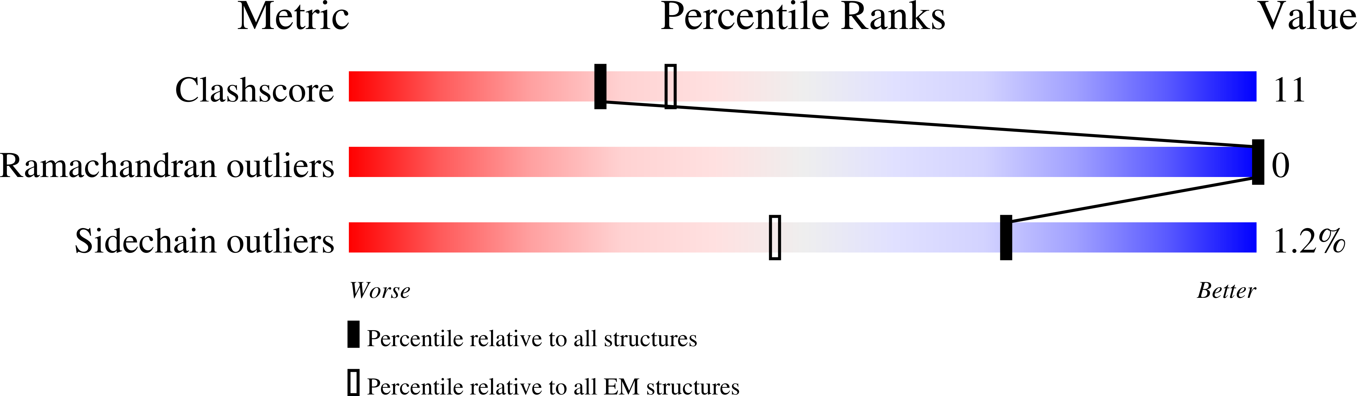
    6X4S

  • PubMed Abstract: 

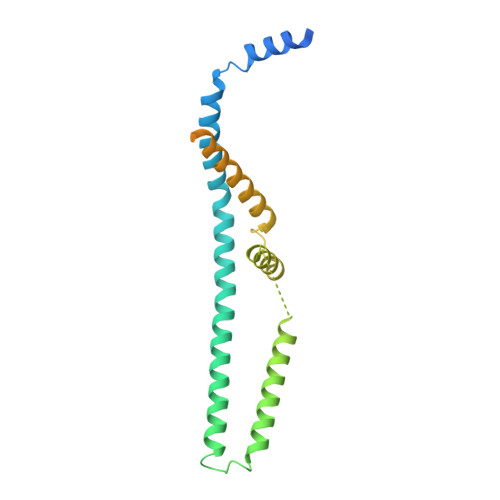
    The proteins MCU and EMRE form the minimal functional unit of the mitochondrial calcium uniporter complex in metazoans, a highly selective and tightly controlled Ca 2+ channel of the inner mitochondrial membrane that regulates cellular metabolism. Here we present functional reconstitution of an MCU-EMRE complex from the red flour beetle, Tribolium castaneum, and a cryo-EM structure of the complex at 3.5 Å resolution. Using a novel assay, we demonstrate robust Ca 2+ uptake into proteoliposomes containing the purified complex. Uptake is dependent on EMRE and also on the mitochondrial lipid cardiolipin. The structure reveals a tetrameric channel with a single ion pore. EMRE is located at the periphery of the transmembrane domain and associates primarily with the first transmembrane helix of MCU. Coiled-coil and juxtamembrane domains within the matrix portion of the complex adopt markedly different conformations than in a structure of a human MCU-EMRE complex, suggesting that the structures represent different conformations of these functionally similar metazoan channels.


  • Organizational Affiliation

    Structural Biology Program, Memorial Sloan Kettering Cancer Center, 1275 York Avenue, New York, NY 10065, USA.


Macromolecules
Find similar proteins by:  (by identity cutoff)  |  3D Structure
Entity ID: 1
MoleculeChains Sequence LengthOrganismDetailsImage
Calcium uniporter protein,Protein EMRE homolog, mitochondrial-like Protein fusion
A, B, C, D, E
A, B, C, D, E, F, G, H
243Tribolium castaneumMutation(s): 0 
Gene Names: TcasGA2_TC013837TcasGA2_TC012057
Membrane Entity: Yes 
UniProt
Find proteins for D6X268 (Tribolium castaneum)
Explore D6X268 
Go to UniProtKB:  D6X268
Find proteins for D6WIX5 (Tribolium castaneum)
Explore D6WIX5 
Go to UniProtKB:  D6WIX5
Entity Groups  
Sequence Clusters30% Identity50% Identity70% Identity90% Identity95% Identity100% Identity
UniProt GroupsD6WIX5D6X268
Sequence Annotations
Expand
  • Reference Sequence
Small Molecules
Ligands 1 Unique
IDChains Name / Formula / InChI Key2D Diagram3D Interactions
CA (Subject of Investigation/LOI)
Query on CA

Download Ideal Coordinates CCD File 
I [auth A]CALCIUM ION
Ca
BHPQYMZQTOCNFJ-UHFFFAOYSA-N
Experimental Data & Validation

Experimental Data

  • Method: ELECTRON MICROSCOPY
  • Resolution: 3.50 Å
  • Aggregation State: PARTICLE 
  • Reconstruction Method: SINGLE PARTICLE 
EM Software:
TaskSoftware PackageVersion
RECONSTRUCTIONRELION3.0
MODEL REFINEMENTPHENIX1.17.1-3660

Structure Validation

View Full Validation Report


Entry History & Funding Information

Deposition Data


Funding OrganizationLocationGrant Number
National Institutes of Health/National Institute of General Medical Sciences (NIH/NIGMS)United StatesR35GM131921
National Institutes of Health/National Cancer Institute (NIH/NCI)United StatesP30CA008748

Revision History  (Full details and data files)

  • Version 1.0: 2020-09-02
    Type: Initial release
  • Version 1.1: 2020-09-30
    Changes: Database references
  • Version 1.2: 2020-10-14
    Changes: Database references
  • Version 1.3: 2024-03-06
    Changes: Data collection, Database references