6WF2

Crystal structure of mouse SCD1 with a diiron center

  • Classification: OXIDOREDUCTASE
  • Organism(s): Mus musculus
  • Expression System: Homo sapiens
  • Mutation(s): No 
  • Membrane Protein: Yes  PDBTMMemProtMDmpstruc

  • Deposited: 2020-04-03 Released: 2020-06-17 
  • Deposition Author(s): Shen, J., Zhou, M.
  • Funding Organization(s): National Institutes of Health/National Institute of Diabetes and Digestive and Kidney Disease (NIH/NIDDK), National Institutes of Health/National Heart, Lung, and Blood Institute (NIH/NHLBI), Cancer Prevention and Research Institute of Texas (CPRIT)

Experimental Data Snapshot

  • Method: X-RAY DIFFRACTION
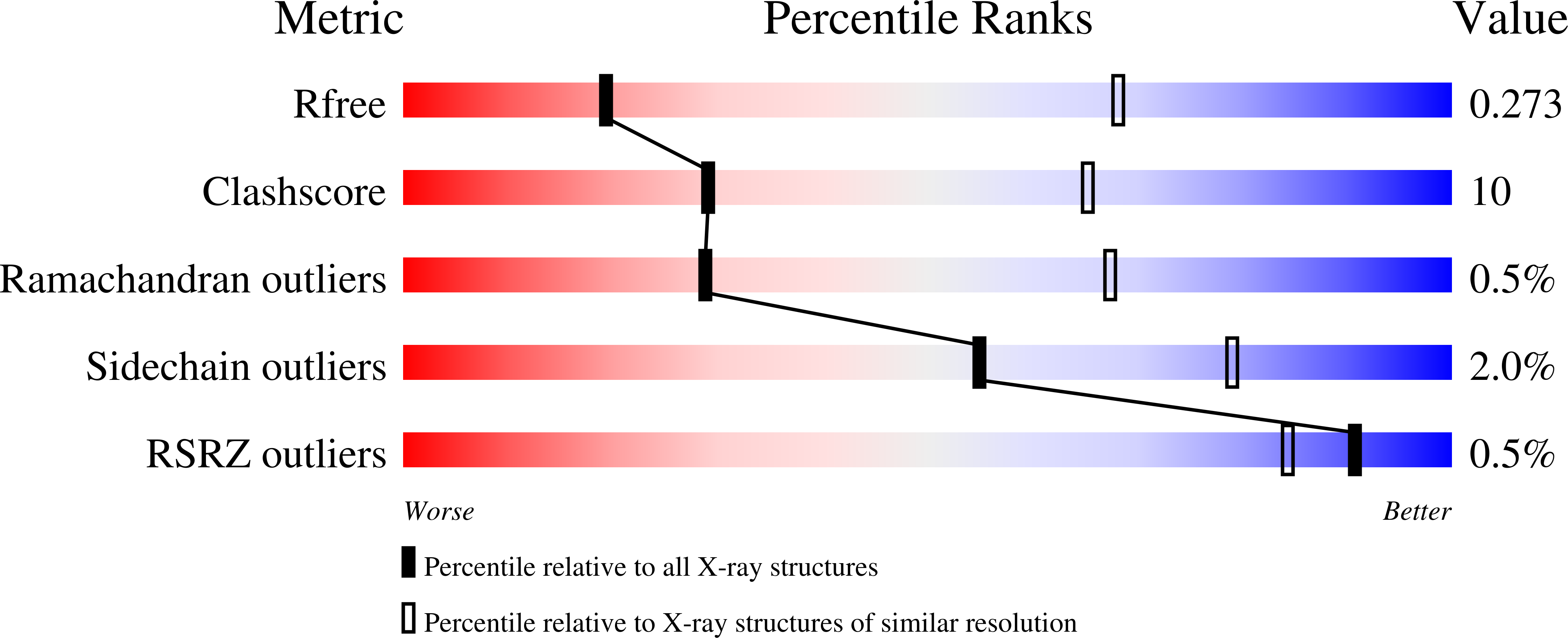
  • Resolution: 3.51 Å
  • R-Value Free: 0.277 
  • R-Value Work: 0.219 
  • R-Value Observed: 0.224 

wwPDB Validation   3D Report Full Report


This is version 1.2 of the entry. See complete history


Literature

Structure and Mechanism of a Unique Diiron Center in Mammalian Stearoyl-CoA Desaturase.

Shen, J.Wu, G.Tsai, A.L.Zhou, M.

(2020) J Mol Biol 432: 5152-5161

  • DOI: https://doi.org/10.1016/j.jmb.2020.05.017
  • Primary Citation of Related Structures:  
    6WF2

  • PubMed Abstract: 

    Stearoyl-CoA desaturase 1 (SCD1) is a membrane-embedded metalloenzyme that catalyzes the formation of a double bond on a saturated acyl-CoA. SCD1 has a diiron center and its proper function requires an electron transport chain composed of NADH (or NADPH), cytochrome b 5 reductase (b 5 R), and cytochrome b 5 (cyt b 5 ). Since SCD1 is a key regulator in fat metabolism and is required for survival of cancer cells, there is intense interest in targeting SCD1 for various metabolic diseases and cancers. Crystal structures of human and mouse SCD1 were reported recently; however, both proteins have two zinc ions instead of two iron ions in the catalytic center, and as a result, the enzymes are inactive. Here we report a general approach for incorporating iron into heterologously expressed proteins in HEK293 cells. We produced mouse SCD1 that contains a diiron center and visualized its diiron center by solving its crystal structure to 3.5 Å. We assembled the entire electron transport chain using the purified soluble domains of cyt b 5 and b 5 R, and the purified mouse SCD1, and we showed that three proteins coordinate to produce proper products. These results established an in vitro system that allows precise perturbations of the electron transport chain for the understanding of the catalytic mechanism in SCD1.


  • Organizational Affiliation

    Verna and Marrs McLean Department of Biochemistry and Molecular Biology, Baylor College of Medicine, One Baylor Plaza, Houston, TX 77030, USA.


Macromolecules
Find similar proteins by:  (by identity cutoff)  |  3D Structure
Entity ID: 1
MoleculeChains Sequence LengthOrganismDetailsImage
Acyl-CoA desaturase 1
A, B
339Mus musculusMutation(s): 0 
Gene Names: Scd1
EC: 1.14.19.1
Membrane Entity: Yes 
UniProt
Find proteins for P13516 (Mus musculus)
Explore P13516 
Go to UniProtKB:  P13516
Entity Groups  
Sequence Clusters30% Identity50% Identity70% Identity90% Identity95% Identity100% Identity
UniProt GroupP13516
Sequence Annotations
Expand
  • Reference Sequence
Small Molecules
Ligands 2 Unique
IDChains Name / Formula / InChI Key2D Diagram3D Interactions
3VV
Query on 3VV

Download Ideal Coordinates CCD File 
E [auth A],
H [auth B]
S-{(3R,5R,9R)-1-[(2R,3S,4R,5R)-5-(6-amino-9H-purin-9-yl)-4-hydroxy-3-(phosphonooxy)tetrahydrofuran-2-yl]-3,5,9-trihydroxy-8,8-dimethyl-3,5-dioxido-10,14-dioxo-2,4,6-trioxa-11,15-diaza-3lambda~5~,5lambda~5~-diphosphaheptadecan-17-yl} (9Z)-octadec-9-enethioate (non-preferred name)
C39 H68 N7 O17 P3 S
XDUHQPOXLUAVEE-BPMMELMSSA-N
FE (Subject of Investigation/LOI)
Query on FE

Download Ideal Coordinates CCD File 
C [auth A],
D [auth A],
F [auth B],
G [auth B]
FE (III) ION
Fe
VTLYFUHAOXGGBS-UHFFFAOYSA-N
Experimental Data & Validation

Experimental Data

  • Method: X-RAY DIFFRACTION
  • Resolution: 3.51 Å
  • R-Value Free: 0.277 
  • R-Value Work: 0.219 
  • R-Value Observed: 0.224 
  • Space Group: P 21 21 21
Unit Cell:
Length ( Å )Angle ( ˚ )
a = 76.647α = 90
b = 113.981β = 90
c = 140.971γ = 90
Software Package:
Software NamePurpose
PHENIXrefinement
XDSdata reduction
XDSdata scaling
PHASERphasing

Structure Validation

View Full Validation Report


Entry History & Funding Information

Deposition Data


Funding OrganizationLocationGrant Number
National Institutes of Health/National Institute of Diabetes and Digestive and Kidney Disease (NIH/NIDDK)United StatesDK122784
National Institutes of Health/National Heart, Lung, and Blood Institute (NIH/NHLBI)United StatesHL086392
Cancer Prevention and Research Institute of Texas (CPRIT)United StatesR1223

Revision History  (Full details and data files)

  • Version 1.0: 2020-06-17
    Type: Initial release
  • Version 1.1: 2020-09-02
    Changes: Database references
  • Version 1.2: 2023-10-18
    Changes: Data collection, Database references, Refinement description