6USC

Structure of Human Intelectin-1 in complex with KO

  • Classification: SUGAR BINDING PROTEIN
  • Organism(s): Homo sapiens
  • Expression System: Homo sapiens
  • Mutation(s): No 

  • Deposited: 2019-10-25 Released: 2020-01-22 
  • Deposition Author(s): Windsor, I.W., Isabella, C.R., Kosma, P., Raines, R.T., Kiessling, L.L.
  • Funding Organization(s): National Institutes of Health/National Institute Of Allergy and Infectious Diseases (NIH/NIAID), National Institutes of Health/National Cancer Institute (NIH/NCI), National Institutes of Health/National Institute of General Medical Sciences (NIH/NIGMS), Austrian Science Fund, National Science Foundation (NSF, United States)

Experimental Data Snapshot

  • Method: X-RAY DIFFRACTION
  • Resolution: 1.59 Å
  • R-Value Free: 0.182 
  • R-Value Work: 0.158 
  • R-Value Observed: 0.159 

wwPDB Validation   3D Report Full Report

Currently 6USC does not have a validation slider image.


This is version 1.3 of the entry. See complete history


Literature

Stereoelectronic Effects Impact Glycan Recognition.

McMahon, C.M.Isabella, C.R.Windsor, I.W.Kosma, P.Raines, R.T.Kiessling, L.L.

(2020) J Am Chem Soc 142: 2386-2395

  • DOI: https://doi.org/10.1021/jacs.9b11699
  • Primary Citation of Related Structures:  
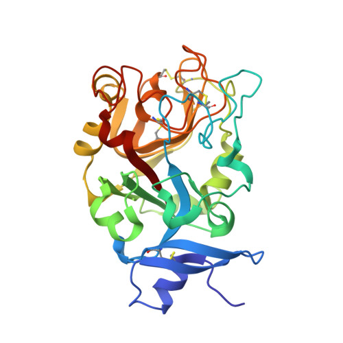
    6USC

  • PubMed Abstract: 

    Recognition of distinct glycans is central to biology, and lectins mediate this function. Lectin glycan preferences are usually centered on specific monosaccharides. In contrast, human intelectin-1 (hItln-1, also known as Omentin-1) is a soluble lectin that binds a range of microbial sugars, including β-d-galactofuranose (β-Gal f ), d-glycerol 1-phosphate, d- glycero -d- talo -oct-2-ulosonic acid (KO), and 3-deoxy-d- manno -oct-2-ulosonic acid (KDO). Though these saccharides differ dramatically in structure, they share a common feature-an exocyclic vicinal diol. How and whether such a small fragment is sufficient for recognition was unclear. We tested several glycans with this epitope and found that l- glycero -α-d- manno -heptose and d- glycero -α-d- manno -heptose possess the critical diol motif yet bind weakly. To better understand hItln-1 recognition, we determined the structure of the hItln-1·KO complex using X-ray crystallography, and our 1.59 Å resolution structure enabled unambiguous assignment of the bound KO conformation. This carbohydrate conformation was present in >97% of the KDO/KO structures in the Protein Data Bank. Bioinformatic analysis revealed that KO and KDO adopt a common conformation, while heptoses prefer different conformers. The preferred conformers of KO and KDO favor hItln-1 engagement, but those of the heptoses do not. Natural bond orbital (NBO) calculations suggest these observed conformations, including the side chain orientations, are stabilized by not only steric but also stereoelectronic effects. Thus, our data highlight a role for stereoelectronic effects in dictating the specificity of glycan recognition by proteins. Finally, our finding that hItln-1 avoids binding prevalent glycans with a terminal 1,2-diol (e.g., N -acetyl-neuraminic acid and l- glycero -α-d- manno -heptose) suggests the lectin has evolved to recognize distinct bacterial species.


  • Organizational Affiliation

    Department of Chemistry , Massachusetts Institute of Technology , Cambridge , Massachusetts 02139 , United States.


Macromolecules
Find similar proteins by:  (by identity cutoff)  |  3D Structure
Entity ID: 1
MoleculeChains Sequence LengthOrganismDetailsImage
Intelectin-1
A, B
279Homo sapiensMutation(s): 0 
Gene Names: ITLN1INTLITLNLFRUNQ640/PRO1270
UniProt & NIH Common Fund Data Resources
Find proteins for Q8WWA0 (Homo sapiens)
Explore Q8WWA0 
Go to UniProtKB:  Q8WWA0
PHAROS:  Q8WWA0
GTEx:  ENSG00000179914 
Entity Groups  
Sequence Clusters30% Identity50% Identity70% Identity90% Identity95% Identity100% Identity
UniProt GroupQ8WWA0
Sequence Annotations
Expand
  • Reference Sequence
Small Molecules
Ligands 3 Unique
IDChains Name / Formula / InChI Key2D Diagram3D Interactions
KO2 (Subject of Investigation/LOI)
Query on KO2

Download Ideal Coordinates CCD File 
F [auth A],
K [auth B]
prop-2-en-1-yl D-glycero-alpha-D-talo-oct-2-ulopyranosidonic acid
C11 H18 O9
MJWRJGHIYDIAQR-RUEXUQCSSA-N
CA
Query on CA

Download Ideal Coordinates CCD File 
C [auth A]
D [auth A]
E [auth A]
H [auth B]
I [auth B]
C [auth A],
D [auth A],
E [auth A],
H [auth B],
I [auth B],
J [auth B]
CALCIUM ION
Ca
BHPQYMZQTOCNFJ-UHFFFAOYSA-N
CL
Query on CL

Download Ideal Coordinates CCD File 
G [auth A]CHLORIDE ION
Cl
VEXZGXHMUGYJMC-UHFFFAOYSA-M
Binding Affinity Annotations 
IDSourceBinding Affinity
KO2 Binding MOAD:  6USC IC50: 7.00e+5 (nM) from 1 assay(s)
Experimental Data & Validation

Experimental Data

  • Method: X-RAY DIFFRACTION
  • Resolution: 1.59 Å
  • R-Value Free: 0.182 
  • R-Value Work: 0.158 
  • R-Value Observed: 0.159 
  • Space Group: P 21 3
Unit Cell:
Length ( Å )Angle ( ˚ )
a = 118.145α = 90
b = 118.145β = 90
c = 118.145γ = 90
Software Package:
Software NamePurpose
PHENIXrefinement
Aimlessdata scaling
PDB_EXTRACTdata extraction
DIALSdata reduction
PHASERphasing

Structure Validation

View Full Validation Report

Currently 6USC does not have a validation slider image.


Entry History & Funding Information

Deposition Data


Funding OrganizationLocationGrant Number
National Institutes of Health/National Institute Of Allergy and Infectious Diseases (NIH/NIAID)United StatesR01AI055258
National Institutes of Health/National Cancer Institute (NIH/NCI)United StatesU01 CA2310789
National Institutes of Health/National Institute of General Medical Sciences (NIH/NIGMS)United StatesR01GM044783
Austrian Science FundAustriaP28826-N28
National Science Foundation (NSF, United States)United States1122374

Revision History  (Full details and data files)

  • Version 1.0: 2020-01-22
    Type: Initial release
  • Version 1.1: 2020-02-19
    Changes: Database references
  • Version 1.2: 2020-07-29
    Type: Remediation
    Reason: Carbohydrate remediation
    Changes: Data collection, Derived calculations
  • Version 1.3: 2023-10-11
    Changes: Data collection, Database references, Refinement description, Structure summary