6UQV

Crystal structure of ChoE, a bacterial acetylcholinesterase from Pseudomonas aeruginosa


Experimental Data Snapshot

  • Method: X-RAY DIFFRACTION
  • Resolution: 1.35 Å
  • R-Value Free: 0.177 
  • R-Value Work: 0.154 
  • R-Value Observed: 0.155 

wwPDB Validation   3D Report Full Report


Ligand Structure Quality Assessment 


This is version 1.2 of the entry. See complete history


Literature

Structural insights into the putative bacterial acetylcholinesterase ChoE and its substrate inhibition mechanism.

Pham, V.D.To, T.A.Gagne-Thivierge, C.Couture, M.Lague, P.Yao, D.Picard, M.E.Lortie, L.A.Attere, S.A.Zhu, X.Levesque, R.C.Charette, S.J.Shi, R.

(2020) J Biol Chem 295: 8708-8724

  • DOI: https://doi.org/10.1074/jbc.RA119.011809
  • Primary Citation of Related Structures:  
    6UQV, 6UQW, 6UQX, 6UQY, 6UQZ, 6UR0, 6UR1

  • PubMed Abstract: 

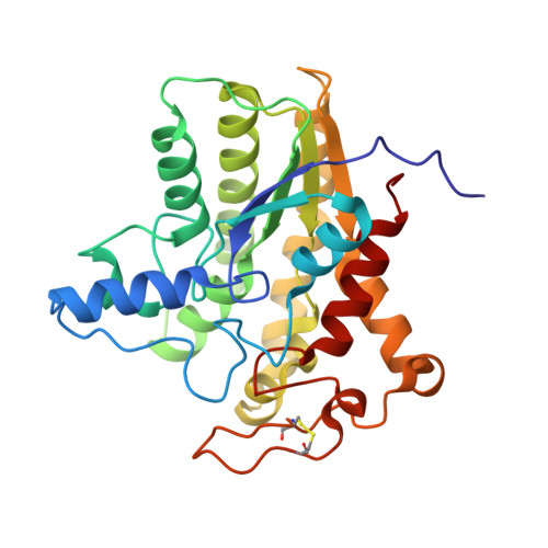
    Mammalian acetylcholinesterase (AChE) is well-studied, being important in both cholinergic brain synapses and the peripheral nervous systems and also a key drug target for many diseases. In contrast, little is known about the structures and molecular mechanism of prokaryotic acetylcholinesterases. We report here the structural and biochemical characterization of ChoE, a putative bacterial acetylcholinesterase from Pseudomonas aeruginosa Analysis of WT and mutant strains indicated that ChoE is indispensable for P. aeruginosa growth with acetylcholine as the sole carbon and nitrogen source. The crystal structure of ChoE at 1.35 Å resolution revealed that this enzyme adopts a typical fold of the SGNH hydrolase family. Although ChoE and eukaryotic AChEs catalyze the same reaction, their overall structures bear no similarities constituting an interesting example of convergent evolution. Among Ser-38, Asp-285, and His-288 of the catalytic triad residues, only Asp-285 was not essential for ChoE activity. Combined with kinetic analyses of WT and mutant proteins, multiple crystal structures of ChoE complexed with substrates, products, or reaction intermediate revealed the structural determinants for substrate recognition, snapshots of the various catalytic steps, and the molecular basis of substrate inhibition at high substrate concentrations. Our results indicate that substrate inhibition in ChoE is due to acetate release being blocked by the binding of a substrate molecule in a nonproductive mode. Because of the distinct overall folds and significant differences of the active site between ChoE and eukaryotic AChEs, these structures will serve as a prototype for other prokaryotic acetylcholinesterases.


  • Organizational Affiliation

    Département de Biochimie, de Microbiologie et de Bio-informatique, Université Laval, Québec, Canada; Institut de Biologie Intégrative et des Systèmes (IBIS), Université Laval, Québec, Canada; PROTEO, the Québec Network for Research on Protein Function, Engineering, and Applications, Québec, Canada.


Macromolecules
Find similar proteins by:  (by identity cutoff)  |  3D Structure
Entity ID: 1
MoleculeChains Sequence LengthOrganismDetailsImage
ChoE287Pseudomonas aeruginosa PAO1Mutation(s): 0 
Gene Names: choEPA4921
EC: 3.1
UniProt
Find proteins for Q9HUP2 (Pseudomonas aeruginosa (strain ATCC 15692 / DSM 22644 / CIP 104116 / JCM 14847 / LMG 12228 / 1C / PRS 101 / PAO1))
Explore Q9HUP2 
Go to UniProtKB:  Q9HUP2
Entity Groups  
Sequence Clusters30% Identity50% Identity70% Identity90% Identity95% Identity100% Identity
UniProt GroupQ9HUP2
Sequence Annotations
Expand
  • Reference Sequence
Small Molecules
Ligands 5 Unique
IDChains Name / Formula / InChI Key2D Diagram3D Interactions
P6G
Query on P6G

Download Ideal Coordinates CCD File 
F [auth A]HEXAETHYLENE GLYCOL
C12 H26 O7
IIRDTKBZINWQAW-UHFFFAOYSA-N
MES (Subject of Investigation/LOI)
Query on MES

Download Ideal Coordinates CCD File 
B [auth A]2-(N-MORPHOLINO)-ETHANESULFONIC ACID
C6 H13 N O4 S
SXGZJKUKBWWHRA-UHFFFAOYSA-N
GOL
Query on GOL

Download Ideal Coordinates CCD File 
C [auth A]GLYCEROL
C3 H8 O3
PEDCQBHIVMGVHV-UHFFFAOYSA-N
BUA
Query on BUA

Download Ideal Coordinates CCD File 
D [auth A]butanoic acid
C4 H8 O2
FERIUCNNQQJTOY-UHFFFAOYSA-N
CL
Query on CL

Download Ideal Coordinates CCD File 
E [auth A]CHLORIDE ION
Cl
VEXZGXHMUGYJMC-UHFFFAOYSA-M
Experimental Data & Validation

Experimental Data

  • Method: X-RAY DIFFRACTION
  • Resolution: 1.35 Å
  • R-Value Free: 0.177 
  • R-Value Work: 0.154 
  • R-Value Observed: 0.155 
  • Space Group: C 2 2 21
Unit Cell:
Length ( Å )Angle ( ˚ )
a = 82.157α = 90
b = 109.673β = 90
c = 84.22γ = 90
Software Package:
Software NamePurpose
SCALAdata scaling
REFMACrefinement
PDB_EXTRACTdata extraction
MOSFLMdata reduction
SHELXDphasing

Structure Validation

View Full Validation Report


Ligand Structure Quality Assessment 


Entry History & Funding Information

Deposition Data


Funding OrganizationLocationGrant Number
Fonds de Recherche du Quebec - Nature et Technologies (FRQNT)Canada183530

Revision History  (Full details and data files)

  • Version 1.0: 2020-05-13
    Type: Initial release
  • Version 1.1: 2020-05-20
    Changes: Database references
  • Version 1.2: 2020-07-08
    Changes: Database references