6UKA

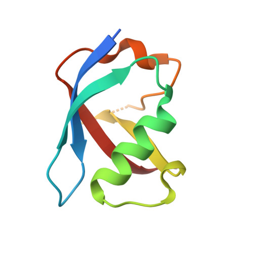
Crystal structure of RHOG and ELMO complex


Experimental Data Snapshot

  • Method: X-RAY DIFFRACTION
  • Resolution: 2.40 Å
  • R-Value Free: 0.237 
  • R-Value Work: 0.186 
  • R-Value Observed: 0.191 

wwPDB Validation   3D Report Full Report

Currently 6UKA does not have a validation slider image.


This is version 1.1 of the entry. See complete history


Literature

Structure of the DOCK2-ELMO1 complex provides insights into regulation of the auto-inhibited state.

Chang, L.Yang, J.Jo, C.H.Boland, A.Zhang, Z.McLaughlin, S.H.Abu-Thuraia, A.Killoran, R.C.Smith, M.J.Cote, J.F.Barford, D.

(2020) Nat Commun 11: 3464-3464

  • DOI: https://doi.org/10.1038/s41467-020-17271-9
  • Primary Citation of Related Structures:  
    6TGB, 6TGC, 6UKA

  • PubMed Abstract: 

    DOCK (dedicator of cytokinesis) proteins are multidomain guanine nucleotide exchange factors (GEFs) for RHO GTPases that regulate intracellular actin dynamics. DOCK proteins share catalytic (DOCK DHR2 ) and membrane-associated (DOCK DHR1 ) domains. The structurally-related DOCK1 and DOCK2 GEFs are specific for RAC, and require ELMO (engulfment and cell motility) proteins for function. The N-terminal RAS-binding domain (RBD) of ELMO (ELMO RBD ) interacts with RHOG to modulate DOCK1/2 activity. Here, we determine the cryo-EM structures of DOCK2-ELMO1 alone, and as a ternary complex with RAC1, together with the crystal structure of a RHOG-ELMO2 RBD complex. The binary DOCK2-ELMO1 complex adopts a closed, auto-inhibited conformation. Relief of auto-inhibition to an active, open state, due to a conformational change of the ELMO1 subunit, exposes binding sites for RAC1 on DOCK2 DHR2 , and RHOG and BAI GPCRs on ELMO1. Our structure explains how up-stream effectors, including DOCK2 and ELMO1 phosphorylation, destabilise the auto-inhibited state to promote an active GEF.


  • Organizational Affiliation

    MRC Laboratory of Molecular Biology, Cambridge, CB2 0QH, UK.


Macromolecules
Find similar proteins by:  (by identity cutoff)  |  3D Structure
Entity ID: 1
MoleculeChains Sequence LengthOrganismDetailsImage
Rho-related GTP-binding protein RhoG191Homo sapiensMutation(s): 0 
Gene Names: RHOGARHG
UniProt & NIH Common Fund Data Resources
Find proteins for P84095 (Homo sapiens)
Explore P84095 
Go to UniProtKB:  P84095
PHAROS:  P84095
GTEx:  ENSG00000177105 
Entity Groups  
Sequence Clusters30% Identity50% Identity70% Identity90% Identity95% Identity100% Identity
UniProt GroupP84095
Sequence Annotations
Expand
  • Reference Sequence
Find similar proteins by:  (by identity cutoff)  |  3D Structure
Entity ID: 2
MoleculeChains Sequence LengthOrganismDetailsImage
Engulfment and cell motility protein 281Mus musculusMutation(s): 0 
Gene Names: Elmo2Kiaa1834
UniProt
Find proteins for Q8BHL5 (Mus musculus)
Explore Q8BHL5 
Go to UniProtKB:  Q8BHL5
Entity Groups  
Sequence Clusters30% Identity50% Identity70% Identity90% Identity95% Identity100% Identity
UniProt GroupQ8BHL5
Sequence Annotations
Expand
  • Reference Sequence
Small Molecules
Ligands 2 Unique
IDChains Name / Formula / InChI Key2D Diagram3D Interactions
GNP (Subject of Investigation/LOI)
Query on GNP

Download Ideal Coordinates CCD File 
C [auth A]PHOSPHOAMINOPHOSPHONIC ACID-GUANYLATE ESTER
C10 H17 N6 O13 P3
UQABYHGXWYXDTK-UUOKFMHZSA-N
MG
Query on MG

Download Ideal Coordinates CCD File 
D [auth A]MAGNESIUM ION
Mg
JLVVSXFLKOJNIY-UHFFFAOYSA-N
Experimental Data & Validation

Experimental Data

  • Method: X-RAY DIFFRACTION
  • Resolution: 2.40 Å
  • R-Value Free: 0.237 
  • R-Value Work: 0.186 
  • R-Value Observed: 0.191 
  • Space Group: C 1 2 1
Unit Cell:
Length ( Å )Angle ( ˚ )
a = 100.279α = 90
b = 40.06β = 119.71
c = 73.93γ = 90
Software Package:
Software NamePurpose
PHENIXphasing
PHENIXrefinement
PROTEUM PLUSdata collection
PROTEUM PLUSdata scaling
PROTEUM PLUSdata reduction

Structure Validation

View Full Validation Report

Currently 6UKA does not have a validation slider image.


Entry History & Funding Information

Deposition Data


Funding OrganizationLocationGrant Number
Canadian Institutes of Health Research (CIHR)Canada--

Revision History  (Full details and data files)

  • Version 1.0: 2020-08-12
    Type: Initial release
  • Version 1.1: 2023-10-11
    Changes: Data collection, Database references, Refinement description