6U8Z

Crystal Structure of Catalytic Domain of Human Phospholipase D1


Experimental Data Snapshot

  • Method: X-RAY DIFFRACTION
  • Resolution: 1.80 Å
  • R-Value Free: 0.214 
  • R-Value Work: 0.174 
  • R-Value Observed: 0.175 

wwPDB Validation   3D Report Full Report


This is version 1.1 of the entry. See complete history


Literature

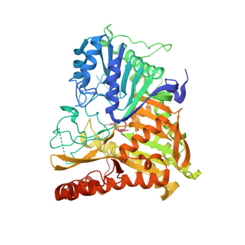
Crystal structure of human PLD1 provides insight into activation by PI(4,5)P2and RhoA.

Bowling, F.Z.Salazar, C.M.Bell, J.A.Huq, T.S.Frohman, M.A.Airola, M.V.

(2020) Nat Chem Biol 16: 400-407

  • DOI: https://doi.org/10.1038/s41589-020-0499-8
  • Primary Citation of Related Structures:  
    6U8Z

  • PubMed Abstract: 

    The signal transduction enzyme phospholipase D1 (PLD1) hydrolyzes phosphatidylcholine to generate the lipid second-messenger phosphatidic acid, which plays roles in disease processes such as thrombosis and cancer. PLD1 is directly and synergistically regulated by protein kinase C, Arf and Rho GTPases, and the membrane lipid phosphatidylinositol-4,5-bisphosphate (PIP 2 ). Here, we present a 1.8 Å-resolution crystal structure of the human PLD1 catalytic domain, which is characterized by a globular fold with a funnel-shaped hydrophobic cavity leading to the active site. Adjacent is a PIP 2 -binding polybasic pocket at the membrane interface that is essential for activity. The C terminus folds into and contributes part of the catalytic pocket, which harbors a phosphohistidine that mimics an intermediate stage of the catalytic cycle. Mapping of PLD1 mutations that disrupt RhoA activation identifies the RhoA-PLD1 binding interface. This structure sheds light on PLD1 regulation by lipid and protein effectors, enabling rationale inhibitor design for this well-studied therapeutic target.


  • Organizational Affiliation

    Department of Biochemistry and Cell Biology, Stony Brook University, Stony Brook, NY, USA.


Macromolecules
Find similar proteins by:  (by identity cutoff)  |  3D Structure
Entity ID: 1
MoleculeChains Sequence LengthOrganismDetailsImage
Phospholipase D1,Phospholipase D1603Homo sapiensMutation(s): 0 
Gene Names: PLD1
EC: 3.1.4.4
Membrane Entity: Yes 
UniProt & NIH Common Fund Data Resources
Find proteins for Q13393 (Homo sapiens)
Explore Q13393 
Go to UniProtKB:  Q13393
PHAROS:  Q13393
GTEx:  ENSG00000075651 
Entity Groups  
Sequence Clusters30% Identity50% Identity70% Identity90% Identity95% Identity100% Identity
UniProt GroupQ13393
Sequence Annotations
Expand
  • Reference Sequence
Small Molecules
Ligands 1 Unique
IDChains Name / Formula / InChI Key2D Diagram3D Interactions
PO3
Query on PO3

Download Ideal Coordinates CCD File 
B [auth A]PHOSPHITE ION
O3 P
AQSJGOWTSHOLKH-UHFFFAOYSA-N
Experimental Data & Validation

Experimental Data

  • Method: X-RAY DIFFRACTION
  • Resolution: 1.80 Å
  • R-Value Free: 0.214 
  • R-Value Work: 0.174 
  • R-Value Observed: 0.175 
  • Space Group: P 41 21 2
Unit Cell:
Length ( Å )Angle ( ˚ )
a = 75.209α = 90
b = 75.209β = 90
c = 239.65γ = 90
Software Package:
Software NamePurpose
xia2data scaling
PHENIXrefinement
PDB_EXTRACTdata extraction
xia2data reduction
AutoSolphasing

Structure Validation

View Full Validation Report


Entry History & Funding Information

Deposition Data


Funding OrganizationLocationGrant Number
National Institutes of Health/National Institute of General Medical Sciences (NIH/NIGMS)United StatesR35GM128666
National Institutes of Health/National Institute of General Medical Sciences (NIH/NIGMS)United StatesT32GM092714

Revision History  (Full details and data files)

  • Version 1.0: 2020-03-18
    Type: Initial release
  • Version 1.1: 2020-04-01
    Changes: Database references