6TWZ

14-3-3 sigma complexed with a phosphorylated 16E6 peptide


Experimental Data Snapshot

  • Method: X-RAY DIFFRACTION
  • Resolution: 2.80 Å
  • R-Value Free: 0.236 
  • R-Value Work: 0.203 
  • R-Value Observed: 0.204 

wwPDB Validation   3D Report Full Report


This is version 1.2 of the entry. See complete history


Literature

Dual Specificity PDZ- and 14-3-3-Binding Motifs: A Structural and Interactomics Study.

Gogl, G.Jane, P.Caillet-Saguy, C.Kostmann, C.Bich, G.Cousido-Siah, A.Nyitray, L.Vincentelli, R.Wolff, N.Nomine, Y.Sluchanko, N.N.Trave, G.

(2020) Structure 28: 747-759.e3

  • DOI: https://doi.org/10.1016/j.str.2020.03.010
  • Primary Citation of Related Structures:  
    6TWQ, 6TWU, 6TWX, 6TWY, 6TWZ

  • PubMed Abstract: 

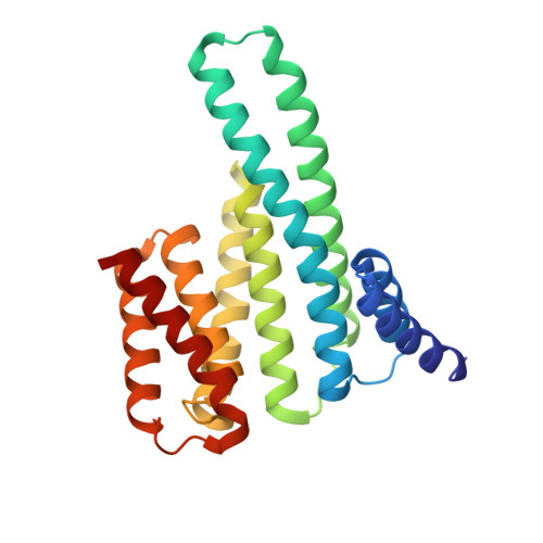
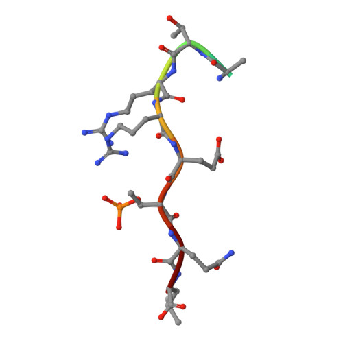
    Protein-protein interaction motifs are often alterable by post-translational modifications. For example, 19% of predicted human PDZ domain-binding motifs (PBMs) have been experimentally proven to be phosphorylated, and up to 82% are theoretically phosphorylatable. Phosphorylation of PBMs may drastically rewire their interactomes, by altering their affinities for PDZ domains and 14-3-3 proteins. The effect of phosphorylation is often analyzed by performing "phosphomimetic" mutations. Here, we focused on the PBMs of HPV16-E6 viral oncoprotein and human RSK1 kinase. We measured the binding affinities of native, phosphorylated, and phosphomimetic variants of both PBMs toward the 266 human PDZ domains. We co-crystallized all the motif variants with a selected PDZ domain to characterize the structural consequence of the different modifications. Finally, we elucidated the structural basis of PBM capture by 14-3-3 proteins. This study provides novel atomic and interactomic insights into phosphorylatable dual specificity motifs and the differential effects of phosphorylation and phosphomimetic approaches.


  • Organizational Affiliation

    Equipe Labellisee Ligue 2015, Department of Integrated Structural Biology, Institut de Genetique et de Biologie Moleculaire et Cellulaire (IGBMC), INSERM U1258/CNRS UMR 7104/Universite de Strasbourg, 1 rue Laurent Fries, BP 10142, 67404 Illkirch, France. Electronic address: goglg@igbmc.fr.


Macromolecules
Find similar proteins by:  (by identity cutoff)  |  3D Structure
Entity ID: 1
MoleculeChains Sequence LengthOrganismDetailsImage
14-3-3 protein sigma
A, B, C, D
234Homo sapiensMutation(s): 3 
Gene Names: SFNHME1
UniProt & NIH Common Fund Data Resources
Find proteins for P31947 (Homo sapiens)
Explore P31947 
Go to UniProtKB:  P31947
PHAROS:  P31947
GTEx:  ENSG00000175793 
Entity Groups  
Sequence Clusters30% Identity50% Identity70% Identity90% Identity95% Identity100% Identity
UniProt GroupP31947
Sequence Annotations
Expand
  • Reference Sequence

Find similar proteins by:  Sequence   |   3D Structure  

Entity ID: 2
MoleculeChains Sequence LengthOrganismDetailsImage
phosphorylated 16E6 peptideE,
F,
G,
H [auth D000]
10Human papillomavirus 16Mutation(s): 0 
Entity Groups  
Sequence Clusters30% Identity50% Identity70% Identity90% Identity95% Identity100% Identity
Sequence Annotations
Expand
  • Reference Sequence
Small Molecules
Modified Residues  1 Unique
IDChains TypeFormula2D DiagramParent
TPO
Query on TPO
E,
F,
G,
H [auth D000]
L-PEPTIDE LINKINGC4 H10 N O6 PTHR
Experimental Data & Validation

Experimental Data

  • Method: X-RAY DIFFRACTION
  • Resolution: 2.80 Å
  • R-Value Free: 0.236 
  • R-Value Work: 0.203 
  • R-Value Observed: 0.204 
  • Space Group: C 1 2 1
Unit Cell:
Length ( Å )Angle ( ˚ )
a = 178.58α = 90
b = 107.42β = 109.289
c = 79.6γ = 90
Software Package:
Software NamePurpose
PHENIXrefinement
PHENIXrefinement
XDSdata reduction
XSCALEdata scaling
PHASERphasing

Structure Validation

View Full Validation Report


Entry History 

Deposition Data

Revision History  (Full details and data files)

  • Version 1.0: 2020-05-06
    Type: Initial release
  • Version 1.1: 2020-07-15
    Changes: Database references
  • Version 1.2: 2024-01-24
    Changes: Data collection, Database references, Refinement description