6TP9

c-type cytochrome NirC


Experimental Data Snapshot

  • Method: X-RAY DIFFRACTION
  • Resolution: 2.19 Å
  • R-Value Free: 0.247 
  • R-Value Work: 0.210 
  • R-Value Observed: 0.212 

wwPDB Validation   3D Report Full Report


Ligand Structure Quality Assessment 


This is version 1.0 of the entry. See complete history


Literature

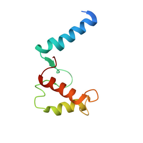
The crystal structure of the heme d1biosynthesis-associated small c-type cytochrome NirC reveals mixed oligomeric states in crystallo.

Klunemann, T.Henke, S.Blankenfeldt, W.

(2020) Acta Crystallogr D Struct Biol 76: 375-384

  • DOI: https://doi.org/10.1107/S2059798320003101
  • Primary Citation of Related Structures:  
    6TP9

  • PubMed Abstract: 

    Monoheme c-type cytochromes are important electron transporters in all domains of life. They possess a common fold hallmarked by three α-helices that surround a covalently attached heme. An intriguing feature of many monoheme c-type cytochromes is their capacity to form oligomers by exchanging at least one of their α-helices, which is often referred to as 3D domain swapping. Here, the crystal structure of NirC, a c-type cytochrome co-encoded with other proteins involved in nitrite reduction by the opportunistic pathogen Pseudomonas aeruginosa, has been determined. The crystals diffracted anisotropically to a maximum resolution of 2.12 Å (spherical resolution of 2.83 Å) and initial phases were obtained by Fe-SAD phasing, revealing the presence of 11 NirC chains in the asymmetric unit. Surprisingly, these protomers arrange into one monomer and two different types of 3D domain-swapped dimers, one of which shows pronounced asymmetry. While the simultaneous observation of monomers and dimers probably reflects the interplay between the high protein concentration required for crystallization and the structural plasticity of monoheme c-type cytochromes, the identification of conserved structural motifs in the monomer together with a comparison with similar proteins may offer new leads to unravel the unknown function of NirC.


  • Organizational Affiliation

    Structure and Function of Proteins, Helmholtz Centre for Infection Research, Inhoffenstrasse 7, 38124 Braunschweig, Germany.


Macromolecules
Find similar proteins by:  (by identity cutoff)  |  3D Structure
Entity ID: 1
MoleculeChains Sequence LengthOrganismDetailsImage
Cytochrome c55X92Pseudomonas aeruginosa PAO1Mutation(s): 1 
Gene Names: nirCPA0517
UniProt
Find proteins for Q51479 (Pseudomonas aeruginosa (strain ATCC 15692 / DSM 22644 / CIP 104116 / JCM 14847 / LMG 12228 / 1C / PRS 101 / PAO1))
Explore Q51479 
Go to UniProtKB:  Q51479
Entity Groups  
Sequence Clusters30% Identity50% Identity70% Identity90% Identity95% Identity100% Identity
UniProt GroupQ51479
Sequence Annotations
Expand
  • Reference Sequence
Small Molecules
Ligands 1 Unique
IDChains Name / Formula / InChI Key2D Diagram3D Interactions
HEC
Query on HEC

Download Ideal Coordinates CCD File 
L [auth A]
M [auth A]
N [auth B]
O [auth B]
P [auth D]
L [auth A],
M [auth A],
N [auth B],
O [auth B],
P [auth D],
Q [auth D],
R [auth E],
S [auth E],
T [auth G],
U [auth H],
V [auth H]
HEME C
C34 H34 Fe N4 O4
HXQIYSLZKNYNMH-LJNAALQVSA-N
Experimental Data & Validation

Experimental Data

  • Method: X-RAY DIFFRACTION
  • Resolution: 2.19 Å
  • R-Value Free: 0.247 
  • R-Value Work: 0.210 
  • R-Value Observed: 0.212 
  • Space Group: P 21 21 21
Unit Cell:
Length ( Å )Angle ( ˚ )
a = 78.017α = 90
b = 81.366β = 90
c = 198.314γ = 90
Software Package:
Software NamePurpose
PHENIXrefinement
Aimlessdata scaling
STARANISOdata scaling
CRANK2phasing
PDB_EXTRACTdata extraction
AutoPROCdata scaling
XDSdata reduction

Structure Validation

View Full Validation Report


Ligand Structure Quality Assessment 


Entry History & Funding Information

Deposition Data


Funding OrganizationLocationGrant Number
German Research Foundation (DFG)GermanyGRK2223/1

Revision History  (Full details and data files)

  • Version 1.0: 2020-04-22
    Type: Initial release