


Crystal structure of the ACVR1 (ALK2) kinase in complex with the compound BI-9564
Williams, E.P., Chen, Z., Burgess-Brown, N., von Delft, F., Arrowsmith, C.H., Edwards, A.M., Bountra, C., Bullock, A.N.To be published.
Experimental Data Snapshot
Entity ID: 1 | |||||
|---|---|---|---|---|---|
| Molecule | Chains | Sequence Length | Organism | Details | Image |
| Activin receptor type I | 301 | Homo sapiens | Mutation(s): 0 Gene Names: ACVR1 EC: 2.7.11.30 | 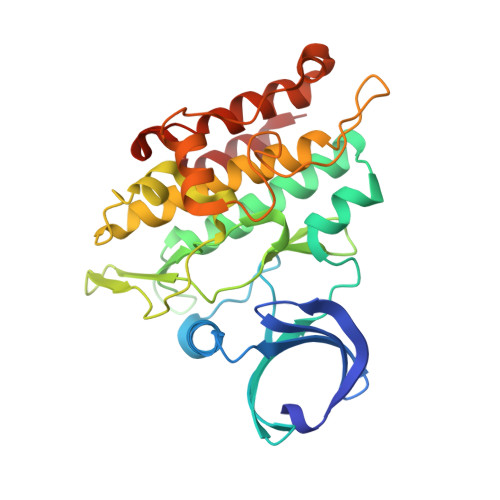 | |
UniProt & NIH Common Fund Data Resources | |||||
Find proteins for Q04771 (Homo sapiens) Explore Q04771 Go to UniProtKB: Q04771 | |||||
PHAROS: Q04771 GTEx: ENSG00000115170 | |||||
Entity Groups | |||||
| Sequence Clusters | 30% Identity50% Identity70% Identity90% Identity95% Identity100% Identity | ||||
| UniProt Group | Q04771 | ||||
Sequence AnnotationsExpand | |||||
| |||||
| Ligands 4 Unique | |||||
|---|---|---|---|---|---|
| ID | Chains | Name / Formula / InChI Key | 2D Diagram | 3D Interactions | |
| 5U6 (Subject of Investigation/LOI) Query on 5U6 | F [auth A] | 4-[4-[(dimethylamino)methyl]-2,5-dimethoxy-phenyl]-2-methyl-2,7-naphthyridin-1-one C20 H23 N3 O3 BJFSUDWKXGMUKA-UHFFFAOYSA-N |  | ||
| SO4 Query on SO4 | G [auth A], H [auth A] | SULFATE ION O4 S QAOWNCQODCNURD-UHFFFAOYSA-L |  | ||
| DIO Query on DIO | B [auth A], C [auth A], D [auth A], E [auth A] | 1,4-DIETHYLENE DIOXIDE C4 H8 O2 RYHBNJHYFVUHQT-UHFFFAOYSA-N |  | ||
| EDO Query on EDO | I [auth A], J [auth A], K [auth A] | 1,2-ETHANEDIOL C2 H6 O2 LYCAIKOWRPUZTN-UHFFFAOYSA-N |  | ||
| Length ( Å ) | Angle ( ˚ ) |
|---|---|
| a = 59.55 | α = 90 |
| b = 86.54 | β = 90 |
| c = 139.599 | γ = 90 |
| Software Name | Purpose |
|---|---|
| PHENIX | refinement |
| PHENIX | refinement |
| iMOSFLM | data reduction |
| Aimless | data scaling |
| PHASER | phasing |
RCSB PDB (citation) is hosted by
RCSB PDB is a member of the

