6TMY

Crystal structure of isoform CBd of the basic phospholipase A2 subunit of crotoxin from Crotalus durissus terrificus


Experimental Data Snapshot

  • Method: X-RAY DIFFRACTION
  • Resolution: 1.80 Å
  • R-Value Free: 0.214 
  • R-Value Work: 0.192 
  • R-Value Observed: 0.192 

Starting Model: experimental
View more details

wwPDB Validation   3D Report Full Report


This is version 1.2 of the entry. See complete history


Literature

Crystal Structure of Isoform CBd of the Basic Phospholipase A 2 Subunit of Crotoxin: Description of the Structural Framework of CB for Interaction with Protein Targets.

Nemecz, D.Ostrowski, M.Ravatin, M.Saul, F.Faure, G.

(2020) Molecules 25

  • DOI: https://doi.org/10.3390/molecules25225290
  • Primary Citation of Related Structures:  
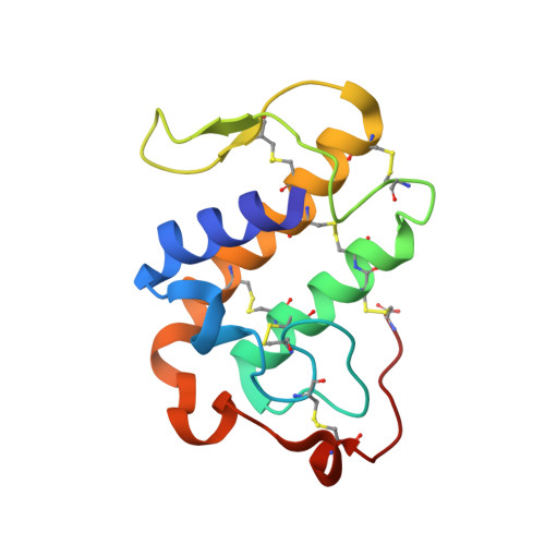
    6TMY

  • PubMed Abstract: 

    Crotoxin, from the venom of the South American rattlesnake Crotalus durissus terrificus, is a potent heterodimeric presynaptic β-neurotoxin that exists in individual snake venom as a mixture of isoforms of a basic phospholipase A 2 (PLA 2 ) subunit (CBa 2 , CBb, CBc, and CBd) and acidic subunit (CA 1-4 ). Specific natural mutations in CB isoforms are implicated in functional differences between crotoxin isoforms. The three-dimensional structure of two individual CB isoforms (CBa 2 , CBc), and one isoform in a crotoxin (CA 2 CBb) complex, have been previously reported. This study concerns CBd, which by interaction with various protein targets exhibits many physiological or pharmacological functions. It binds with high affinity to presynaptic receptors showing neurotoxicity, but also interacts with human coagulation factor Xa (hFXa), exhibiting anticoagulant effect, and acts as a positive allosteric modulator and corrector of mutated chloride channel, cystic fibrosis transmembrane conductance regulator (CFTR), implicated in cystic fibrosis. Thus, CBd represents a novel family of agents that have potential in identifying new drug leads related to anticoagulant and anti-cystic fibrosis function. We determined here the X-ray structure of CBd and compare it with the three other natural isoforms of CB. The structural role of specific amino acid variations between CB isoforms are analyzed and the structural framework of CB for interaction with protein targets is described.


  • Organizational Affiliation

    Institut Pasteur, Récepteurs-Canaux, CNRS UMR 3571, Département de Neuroscience, 25, rue du Dr. Roux, F-75015 Paris, France.


Macromolecules
Find similar proteins by:  (by identity cutoff)  |  3D Structure
Entity ID: 1
MoleculeChains Sequence LengthOrganismDetailsImage
Phospholipase A2 crotoxin basic subunit CBc
A, B, C, D, E
A, B, C, D, E, F
122Crotalus durissus terrificusMutation(s): 0 
EC: 3.1.1.4
UniProt
Find proteins for C0HM14 (Crotalus durissus terrificus)
Explore C0HM14 
Go to UniProtKB:  C0HM14
Entity Groups  
Sequence Clusters30% Identity50% Identity70% Identity90% Identity95% Identity100% Identity
UniProt GroupC0HM14
Sequence Annotations
Expand
  • Reference Sequence
Small Molecules
Ligands 3 Unique
IDChains Name / Formula / InChI Key2D Diagram3D Interactions
PG4
Query on PG4

Download Ideal Coordinates CCD File 
BA [auth E]
DA [auth E]
H [auth A]
HA [auth F]
I [auth A]
BA [auth E],
DA [auth E],
H [auth A],
HA [auth F],
I [auth A],
IA [auth F],
JA [auth F],
O [auth B],
P [auth B],
S [auth C],
T [auth C],
U [auth C],
Y [auth D]
TETRAETHYLENE GLYCOL
C8 H18 O5
UWHCKJMYHZGTIT-UHFFFAOYSA-N
CL
Query on CL

Download Ideal Coordinates CCD File 
EA [auth E]
J [auth A]
K [auth A]
KA [auth F]
L [auth A]
EA [auth E],
J [auth A],
K [auth A],
KA [auth F],
L [auth A],
V [auth C],
Z [auth D]
CHLORIDE ION
Cl
VEXZGXHMUGYJMC-UHFFFAOYSA-M
NA
Query on NA

Download Ideal Coordinates CCD File 
AA [auth D]
CA [auth E]
FA [auth E]
G [auth A]
GA [auth F]
AA [auth D],
CA [auth E],
FA [auth E],
G [auth A],
GA [auth F],
LA [auth F],
M [auth A],
N [auth B],
Q [auth B],
R [auth C],
W [auth C],
X [auth D]
SODIUM ION
Na
FKNQFGJONOIPTF-UHFFFAOYSA-N
Experimental Data & Validation

Experimental Data

  • Method: X-RAY DIFFRACTION
  • Resolution: 1.80 Å
  • R-Value Free: 0.214 
  • R-Value Work: 0.192 
  • R-Value Observed: 0.192 
  • Space Group: C 1 2 1
Unit Cell:
Length ( Å )Angle ( ˚ )
a = 149.626α = 90
b = 75.592β = 121.85
c = 109.012γ = 90
Software Package:
Software NamePurpose
BUSTERrefinement
PDB_EXTRACTdata extraction
XDSdata reduction
Aimlessdata scaling
PHASERphasing

Structure Validation

View Full Validation Report


Entry History & Funding Information

Deposition Data


Funding OrganizationLocationGrant Number
Other governmentPolandPOKL.04.01.01-00-081/10

Revision History  (Full details and data files)

  • Version 1.0: 2020-12-02
    Type: Initial release
  • Version 1.1: 2020-12-09
    Changes: Derived calculations
  • Version 1.2: 2024-01-24
    Changes: Data collection, Database references, Refinement description