6TAE

Neutron structure of ferric ascorbate peroxidase


Experimental Data Snapshot

  • Method: X-RAY DIFFRACTION
  • Resolution: 1.90 Å
  • R-Value Free: 0.189 
  • R-Value Work: 0.144 
  • R-Value Observed: 0.147 

  • Method: NEUTRON DIFFRACTION
  • Resolution: 2.22 Å
  • R-Value Free: 0.308 
  • R-Value Work: 0.276 
  • R-Value Observed: 0.278 

wwPDB Validation   3D Report Full Report


This is version 1.1 of the entry. See complete history


Literature

Visualizing the protons in a metalloenzyme electron proton transfer pathway.

Kwon, H.Basran, J.Devos, J.M.Suardiaz, R.van der Kamp, M.W.Mulholland, A.J.Schrader, T.E.Ostermann, A.Blakeley, M.P.Moody, P.C.E.Raven, E.L.

(2020) Proc Natl Acad Sci U S A 117: 6484-6490

  • DOI: https://doi.org/10.1073/pnas.1918936117
  • Primary Citation of Related Structures:  
    6TAE, 6XV4

  • PubMed Abstract: 

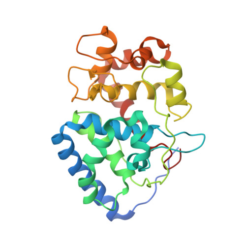
    In redox metalloenzymes, the process of electron transfer often involves the concerted movement of a proton. These processes are referred to as proton-coupled electron transfer, and they underpin a wide variety of biological processes, including respiration, energy conversion, photosynthesis, and metalloenzyme catalysis. The mechanisms of proton delivery are incompletely understood, in part due to an absence of information on exact proton locations and hydrogen bonding structures in a bona fide metalloenzyme proton pathway. Here, we present a 2.1-Å neutron crystal structure of the complex formed between a redox metalloenzyme (ascorbate peroxidase) and its reducing substrate (ascorbate). In the neutron structure of the complex, the protonation states of the electron/proton donor (ascorbate) and all of the residues involved in the electron/proton transfer pathway are directly observed. This information sheds light on possible proton movements during heme-catalyzed oxygen activation, as well as on ascorbate oxidation.


  • Organizational Affiliation

    School of Chemistry, University of Bristol, Bristol BS8 1TS, United Kingdom.


Macromolecules
Find similar proteins by:  (by identity cutoff)  |  3D Structure
Entity ID: 1
MoleculeChains Sequence LengthOrganismDetailsImage
Ascorbate peroxidase261Glycine maxMutation(s): 0 
Gene Names: apx1GLYMA_U021900
EC: 1.11.1.11
UniProt
Find proteins for Q43758 (Glycine max)
Explore Q43758 
Go to UniProtKB:  Q43758
Entity Groups  
Sequence Clusters30% Identity50% Identity70% Identity90% Identity95% Identity100% Identity
UniProt GroupQ43758
Sequence Annotations
Expand
  • Reference Sequence
Small Molecules
Ligands 2 Unique
IDChains Name / Formula / InChI Key2D Diagram3D Interactions
HEM
Query on HEM

Download Ideal Coordinates CCD File 
B [auth A]PROTOPORPHYRIN IX CONTAINING FE
C34 H32 Fe N4 O4
KABFMIBPWCXCRK-RGGAHWMASA-L
SO4
Query on SO4

Download Ideal Coordinates CCD File 
C [auth A]SULFATE ION
O4 S
QAOWNCQODCNURD-UHFFFAOYSA-L
Experimental Data & Validation

Experimental Data

  • Method: X-RAY DIFFRACTION
  • Resolution: 1.90 Å
  • R-Value Free: 0.189 
  • R-Value Work: 0.144 
  • R-Value Observed: 0.147 
  • Space Group: P 42 21 2
  • Method: NEUTRON DIFFRACTION
  • Resolution: 2.22 Å
  • R-Value Free: 0.308 
  • R-Value Work: 0.276 
  • R-Value Observed: 0.278 
  • Space Group: P 42 21 2
Unit Cell:
Length ( Å )Angle ( ˚ )
a = 82.904α = 90
b = 82.904β = 90
c = 75.868γ = 90
Software Package:
Software NamePurpose
PHENIXrefinement
PDB_EXTRACTdata extraction

Structure Validation

View Full Validation Report


Entry History & Funding Information

Deposition Data


Funding OrganizationLocationGrant Number
Biotechnology and Biological Sciences Research CouncilUnited KingdomBB/N015940/1

Revision History  (Full details and data files)

  • Version 1.0: 2020-03-18
    Type: Initial release
  • Version 1.1: 2020-04-08
    Changes: Database references