6SW2

Crystal Structure of P. aeruginosa PqsL in complex with 2-aminobenzoylacetate


Experimental Data Snapshot

  • Method: X-RAY DIFFRACTION
  • Resolution: 1.70 Å
  • R-Value Free: 0.217 
  • R-Value Work: 0.173 
  • R-Value Observed: 0.175 

wwPDB Validation   3D Report Full Report


Ligand Structure Quality Assessment 


This is version 1.2 of the entry. See complete history


Literature

Photoinduced monooxygenation involving NAD(P)H-FAD sequential single-electron transfer.

Ernst, S.Rovida, S.Mattevi, A.Fetzner, S.Drees, S.L.

(2020) Nat Commun 11: 2600-2600

  • DOI: https://doi.org/10.1038/s41467-020-16450-y
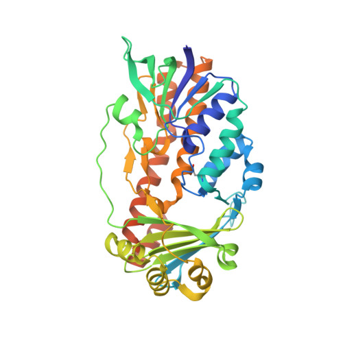
  • Primary Citation of Related Structures:  
    6SW1, 6SW2

  • PubMed Abstract: 

    Light-dependent or light-stimulated catalysis provides a multitude of perspectives for implementation in technological or biomedical applications. Despite substantial progress made in the field of photobiocatalysis, the number of usable light-responsive enzymes is still very limited. Flavoproteins have exceptional potential for photocatalytic applications because the name-giving cofactor intrinsically features light-dependent reactivity, undergoing photoreduction with a variety of organic electron donors. However, in the vast majority of these enzymes, photoreactivity of the enzyme-bound flavin is limited or even suppressed. Here, we present a flavoprotein monooxygenase in which catalytic activity is controllable by blue light illumination. The reaction depends on the presence of nicotinamide nucleotide-type electron donors, which do not support the reaction in the absence of light. Employing various experimental approaches, we demonstrate that catalysis depends on a protein-mediated photoreduction of the flavin cofactor, which proceeds via a radical mechanism and a transient semiquinone intermediate.


  • Organizational Affiliation

    Institute for Molecular Microbiology and Biotechnology, University of Münster, Corrensstr. 3, 48149, Münster, Germany.


Macromolecules
Find similar proteins by:  (by identity cutoff)  |  3D Structure
Entity ID: 1
MoleculeChains Sequence LengthOrganismDetailsImage
Probable FAD-dependent monooxygenase398Pseudomonas aeruginosa PAO1Mutation(s): 0 
Gene Names: pqsLPA4190
UniProt
Find proteins for Q9HWJ1 (Pseudomonas aeruginosa (strain ATCC 15692 / DSM 22644 / CIP 104116 / JCM 14847 / LMG 12228 / 1C / PRS 101 / PAO1))
Explore Q9HWJ1 
Go to UniProtKB:  Q9HWJ1
Entity Groups  
Sequence Clusters30% Identity50% Identity70% Identity90% Identity95% Identity100% Identity
UniProt GroupQ9HWJ1
Sequence Annotations
Expand
  • Reference Sequence
Small Molecules
Ligands 6 Unique
IDChains Name / Formula / InChI Key2D Diagram3D Interactions
FAD (Subject of Investigation/LOI)
Query on FAD

Download Ideal Coordinates CCD File 
B [auth A]FLAVIN-ADENINE DINUCLEOTIDE
C27 H33 N9 O15 P2
VWWQXMAJTJZDQX-UYBVJOGSSA-N
MES
Query on MES

Download Ideal Coordinates CCD File 
F [auth A]2-(N-MORPHOLINO)-ETHANESULFONIC ACID
C6 H13 N O4 S
SXGZJKUKBWWHRA-UHFFFAOYSA-N
61M (Subject of Investigation/LOI)
Query on 61M

Download Ideal Coordinates CCD File 
C [auth A]3-(2-aminophenyl)-3-oxopropanoic acid
C9 H9 N O3
POAXUNDIOGWQOC-UHFFFAOYSA-N
PGE (Subject of Investigation/LOI)
Query on PGE

Download Ideal Coordinates CCD File 
D [auth A],
I [auth A],
J [auth A],
K [auth A]
TRIETHYLENE GLYCOL
C6 H14 O4
ZIBGPFATKBEMQZ-UHFFFAOYSA-N
PEG
Query on PEG

Download Ideal Coordinates CCD File 
G [auth A],
H [auth A]
DI(HYDROXYETHYL)ETHER
C4 H10 O3
MTHSVFCYNBDYFN-UHFFFAOYSA-N
PO4
Query on PO4

Download Ideal Coordinates CCD File 
E [auth A]PHOSPHATE ION
O4 P
NBIIXXVUZAFLBC-UHFFFAOYSA-K
Modified Residues  1 Unique
IDChains TypeFormula2D DiagramParent
CSO
Query on CSO
A
L-PEPTIDE LINKINGC3 H7 N O3 SCYS
Binding Affinity Annotations 
IDSourceBinding Affinity
61M Binding MOAD:  6SW2 Kd: 3.84e+4 (nM) from 1 assay(s)
FAD Binding MOAD:  6SW2 Kd: 259.8 (nM) from 1 assay(s)
Experimental Data & Validation

Experimental Data

  • Method: X-RAY DIFFRACTION
  • Resolution: 1.70 Å
  • R-Value Free: 0.217 
  • R-Value Work: 0.173 
  • R-Value Observed: 0.175 
  • Space Group: P 21 21 21
Unit Cell:
Length ( Å )Angle ( ˚ )
a = 47.079α = 90
b = 63.984β = 90
c = 128.101γ = 90
Software Package:
Software NamePurpose
XDSdata reduction
Aimlessdata scaling
PHASERphasing
REFMACrefinement
PDB_EXTRACTdata extraction

Structure Validation

View Full Validation Report


Ligand Structure Quality Assessment 


Entry History 

Deposition Data

Revision History  (Full details and data files)

  • Version 1.0: 2020-06-03
    Type: Initial release
  • Version 1.1: 2020-06-10
    Changes: Database references
  • Version 1.2: 2024-01-24
    Changes: Data collection, Database references, Refinement description