6SOY

Trypanosoma brucei transferrin receptor in complex with human transferrin


Experimental Data Snapshot

  • Method: X-RAY DIFFRACTION
  • Resolution: 2.75 Å
  • R-Value Free: 0.233 
  • R-Value Work: 0.183 
  • R-Value Observed: 0.185 

wwPDB Validation   3D Report Full Report


Ligand Structure Quality Assessment 


This is version 1.2 of the entry. See complete history


Literature

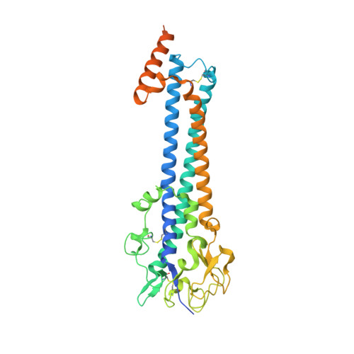
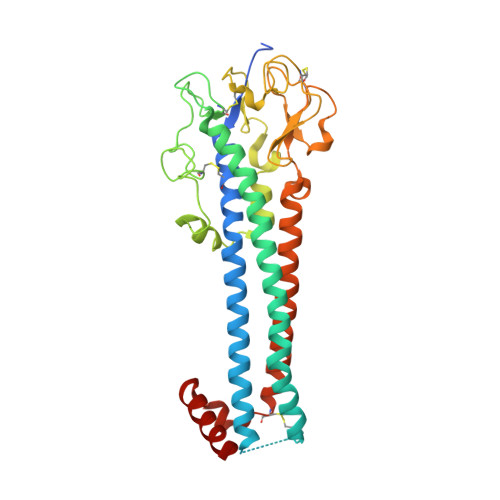
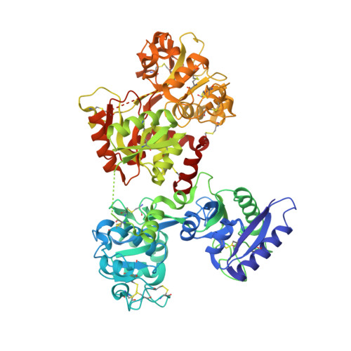
Structure of the trypanosome transferrin receptor reveals mechanisms of ligand recognition and immune evasion.

Trevor, C.E.Gonzalez-Munoz, A.L.Macleod, O.J.S.Woodcock, P.G.Rust, S.Vaughan, T.J.Garman, E.F.Minter, R.Carrington, M.Higgins, M.K.

(2019) Nat Microbiol 4: 2074-2081

  • DOI: https://doi.org/10.1038/s41564-019-0589-0
  • Primary Citation of Related Structures:  
    6SOY, 6SOZ

  • PubMed Abstract: 

    To maintain prolonged infection of mammals, African trypanosomes have evolved remarkable surface coats and a system of antigenic variation 1 . Within these coats are receptors for macromolecular nutrients such as transferrin 2,3 . These must be accessible to their ligands but must not confer susceptibility to immunoglobulin-mediated attack. Trypanosomes have a wide host range and their receptors must also bind ligands from diverse species. To understand how these requirements are achieved, in the context of transferrin uptake, we determined the structure of a Trypanosoma brucei transferrin receptor in complex with human transferrin, showing how this heterodimeric receptor presents a large asymmetric ligand-binding platform. The trypanosome genome contains a family of around 14 transferrin receptors 4 , which has been proposed to allow binding to transferrin from different mammalian hosts 5,6 . However, we find that a single receptor can bind transferrin from a broad range of mammals, indicating that receptor variation is unlikely to be necessary for promiscuity of host infection. In contrast, polymorphic sites and N-linked glycans are preferentially found in exposed positions on the receptor surface, not contacting transferrin, suggesting that transferrin receptor diversification is driven by a need for antigenic variation in the receptor to prolong survival in a host.


  • Organizational Affiliation

    Department of Biochemistry, University of Cambridge, Cambridge, UK.


Macromolecules
Find similar proteins by:  (by identity cutoff)  |  3D Structure
Entity ID: 1
MoleculeChains Sequence LengthOrganismDetailsImage
ESAG6, subunit of heterodimeric transferrin receptor399Trypanosoma bruceiMutation(s): 0 
Gene Names: 13J3.10
UniProt
Find proteins for Q8WPU1 (Trypanosoma brucei brucei)
Explore Q8WPU1 
Go to UniProtKB:  Q8WPU1
Entity Groups  
Sequence Clusters30% Identity50% Identity70% Identity90% Identity95% Identity100% Identity
UniProt GroupQ8WPU1
Sequence Annotations
Expand
  • Reference Sequence
Find similar proteins by:  (by identity cutoff)  |  3D Structure
Entity ID: 2
MoleculeChains Sequence LengthOrganismDetailsImage
ESAG7, subunit of heterodimeric transferrin receptor338Trypanosoma bruceiMutation(s): 0 
Gene Names: 13J3.09
UniProt
Find proteins for Q8WPU2 (Trypanosoma brucei brucei)
Explore Q8WPU2 
Go to UniProtKB:  Q8WPU2
Entity Groups  
Sequence Clusters30% Identity50% Identity70% Identity90% Identity95% Identity100% Identity
UniProt GroupQ8WPU2
Sequence Annotations
Expand
  • Reference Sequence
Find similar proteins by:  (by identity cutoff)  |  3D Structure
Entity ID: 3
MoleculeChains Sequence LengthOrganismDetailsImage
Serotransferrin677Homo sapiensMutation(s): 0 
Gene Names: TFPRO1400
UniProt & NIH Common Fund Data Resources
Find proteins for P02787 (Homo sapiens)
Explore P02787 
Go to UniProtKB:  P02787
PHAROS:  P02787
GTEx:  ENSG00000091513 
Entity Groups  
Sequence Clusters30% Identity50% Identity70% Identity90% Identity95% Identity100% Identity
UniProt GroupP02787
Sequence Annotations
Expand
  • Reference Sequence
Small Molecules
Ligands 2 Unique
IDChains Name / Formula / InChI Key2D Diagram3D Interactions
NAG
Query on NAG

Download Ideal Coordinates CCD File 
E [auth C]2-acetamido-2-deoxy-beta-D-glucopyranose
C8 H15 N O6
OVRNDRQMDRJTHS-FMDGEEDCSA-N
FE
Query on FE

Download Ideal Coordinates CCD File 
D [auth C]FE (III) ION
Fe
VTLYFUHAOXGGBS-UHFFFAOYSA-N
Experimental Data & Validation

Experimental Data

  • Method: X-RAY DIFFRACTION
  • Resolution: 2.75 Å
  • R-Value Free: 0.233 
  • R-Value Work: 0.183 
  • R-Value Observed: 0.185 
  • Space Group: C 1 2 1
Unit Cell:
Length ( Å )Angle ( ˚ )
a = 163.49α = 90
b = 108.11β = 128.74
c = 115γ = 90
Software Package:
Software NamePurpose
BUSTERrefinement
PDB_EXTRACTdata extraction
XDSdata reduction
XDSdata scaling
PHASERphasing

Structure Validation

View Full Validation Report


Ligand Structure Quality Assessment 


Entry History & Funding Information

Deposition Data


Funding OrganizationLocationGrant Number
Medical Research Council (United Kingdom)MR/R001138/1
Biotechnology and Biological Sciences Research Council--
Wellcome Trust--
Engineering and Physical Sciences Research CouncilNS/A000059/1

Revision History  (Full details and data files)

  • Version 1.0: 2019-11-06
    Type: Initial release
  • Version 1.1: 2019-12-04
    Changes: Database references
  • Version 1.2: 2020-07-29
    Type: Remediation
    Reason: Carbohydrate remediation
    Changes: Data collection, Derived calculations, Structure summary