


Crystal Structure of the Homospermidine Synthase (HSS) variant N135F from Blastochloris viridis in Complex with NAD
Scheidig, A.J., Helfrich, F.To be published.
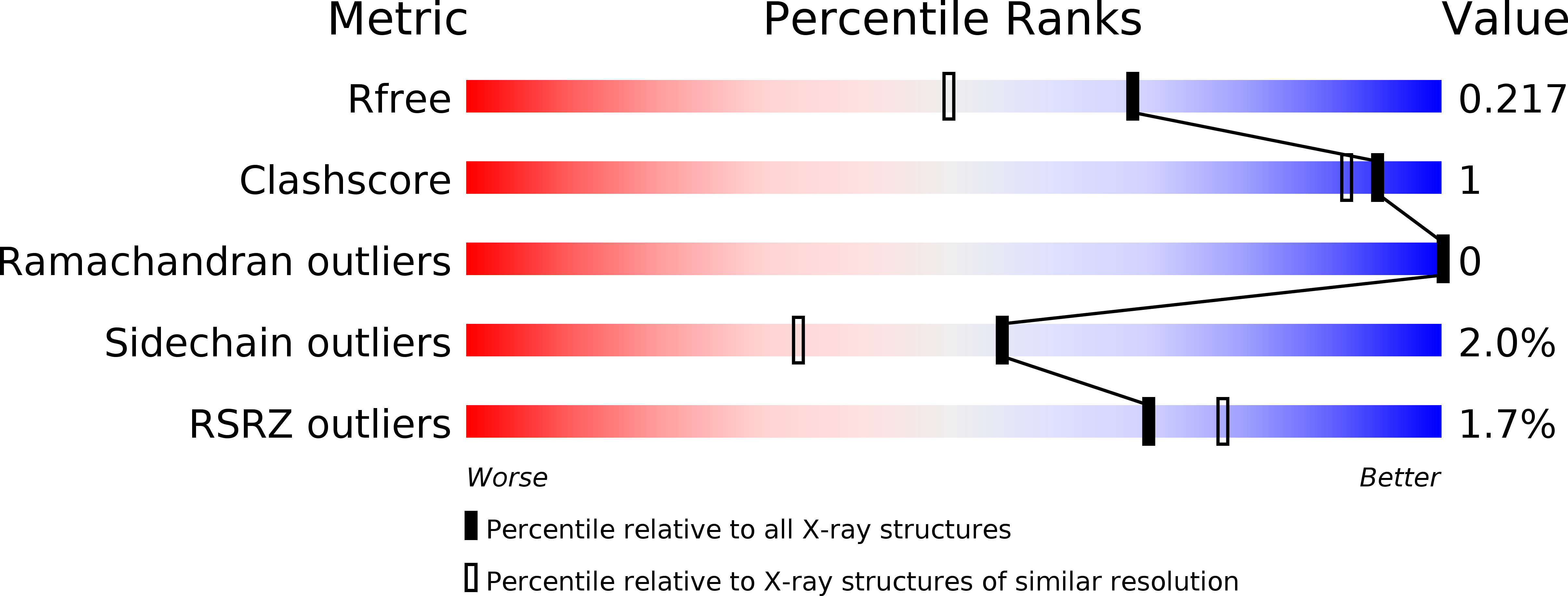
Experimental Data Snapshot
Entity ID: 1 | |||||
|---|---|---|---|---|---|
| Molecule | Chains | Sequence Length | Organism | Details | Image |
| Homospermidine synthase | 480 | Blastochloris viridis | Mutation(s): 1 Gene Names: hss EC: 2.5.1.44 |  | |
UniProt | |||||
Find proteins for O32323 (Blastochloris viridis) Explore O32323 Go to UniProtKB: O32323 | |||||
Entity Groups | |||||
| Sequence Clusters | 30% Identity50% Identity70% Identity90% Identity95% Identity100% Identity | ||||
| UniProt Group | O32323 | ||||
Sequence AnnotationsExpand | |||||
| |||||
| Ligands 1 Unique | |||||
|---|---|---|---|---|---|
| ID | Chains | Name / Formula / InChI Key | 2D Diagram | 3D Interactions | |
| NAD (Subject of Investigation/LOI) Query on NAD | C [auth A], D [auth B] | NICOTINAMIDE-ADENINE-DINUCLEOTIDE C21 H27 N7 O14 P2 BAWFJGJZGIEFAR-NNYOXOHSSA-N |  | ||
| Length ( Å ) | Angle ( ˚ ) |
|---|---|
| a = 60.703 | α = 90 |
| b = 110.401 | β = 90 |
| c = 159.541 | γ = 90 |
| Software Name | Purpose |
|---|---|
| PHENIX | refinement |
| XDS | data reduction |
| Aimless | data scaling |
| PHENIX | phasing |
RCSB PDB (citation) is hosted by
RCSB PDB is a member of the

