6S07

Structure of formylglycine-generating enzyme at 1.04 A in complex with copper and substrate reveals an acidic pocket for binding and acti-vation of molecular oxygen.


Experimental Data Snapshot

  • Method: X-RAY DIFFRACTION
  • Resolution: 1.04 Å
  • R-Value Free: 0.180 
  • R-Value Work: 0.173 
  • R-Value Observed: 0.173 

wwPDB Validation   3D Report Full Report


This is version 2.1 of the entry. See complete history


Literature

Structure of formylglycine-generating enzyme in complex with copper and a substrate reveals an acidic pocket for binding and activation of molecular oxygen.

Miarzlou, D.A.Leisinger, F.Joss, D.Haussinger, D.Seebeck, F.P.

(2019) Chem Sci 10: 7049-7058

  • DOI: https://doi.org/10.1039/c9sc01723b
  • Primary Citation of Related Structures:  
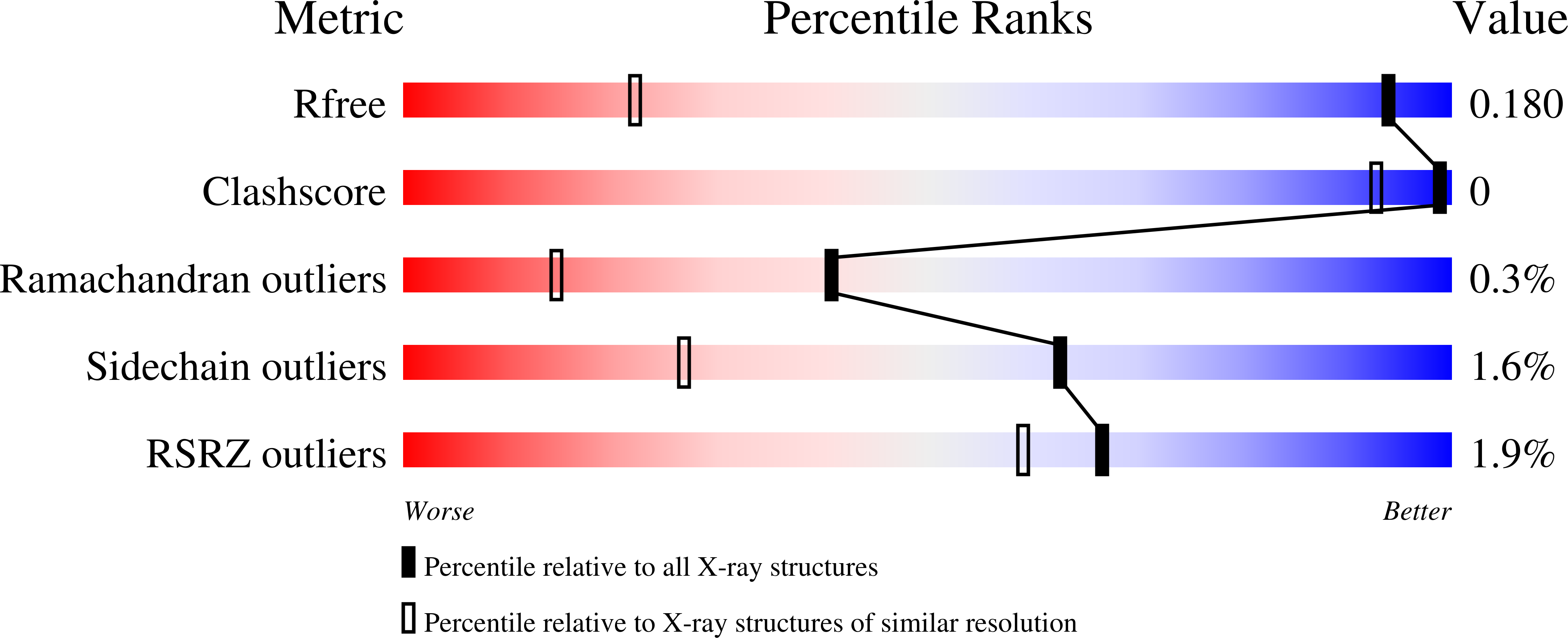
    6S07

  • PubMed Abstract: 

    The formylglycine generating enzyme (FGE) catalyzes oxidative conversion of specific peptidyl-cysteine residues to formylglycine. FGE mediates O 2 -activation and hydrogen-atom abstraction in an active site that contains Cu(i) coordinated to two cysteine residues. Similar coordination geometries are common among copper-sensing transcription factors and copper-chaperone but are unprecedented among copper-dependent oxidases. To examine the mechanism of this unusual catalyst we determined the 1.04 Å structure of FGE from Thermomonospora curvata in complex with copper and a cysteine-containing peptide substrate. This structure unveils a network of four crystallographic waters and two active site residues that form a highly acidic O 2 -binding pocket juxtaposed to the trigonal planar tris-cysteine coordinated Cu(i) center. Comparison with structures of FGE in complex with Ag(i) and Cd(ii) combined with evidence from NMR spectroscopy and kinetic observations highlight several structural changes that are induced by substrate binding and prime the enzyme for O 2 -binding and subsequent activation.


  • Organizational Affiliation

    Department of Chemistry , University of Basel , Mattenstrasse 24a , Basel 4002 , Switzerland . Email: florian.seebeck@unibas.ch ; Tel: +41 612071143.


Macromolecules
Find similar proteins by:  (by identity cutoff)  |  3D Structure
Entity ID: 1
MoleculeChains Sequence LengthOrganismDetailsImage
Formylglycine-generating enzyme303Thermomonospora curvata DSM 43183Mutation(s): 0 
Gene Names: Tcur_4811
EC: 1.8.3.7
UniProt
Find proteins for D1A7C3 (Thermomonospora curvata (strain ATCC 19995 / DSM 43183 / JCM 3096 / KCTC 9072 / NBRC 15933 / NCIMB 10081 / Henssen B9))
Explore D1A7C3 
Go to UniProtKB:  D1A7C3
Entity Groups  
Sequence Clusters30% Identity50% Identity70% Identity90% Identity95% Identity100% Identity
UniProt GroupD1A7C3
Sequence Annotations
Expand
  • Reference Sequence

Find similar proteins by:  Sequence   |   3D Structure  

Entity ID: 2
MoleculeChains Sequence LengthOrganismDetailsImage
Abz-ALA-THR-THR-PRO-LEU-CYS-GLY-PRO-SER-ARG-ALA-SER-ILE-LEU-SER-GLY-ARGB [auth C]17Thermomonospora curvata DSM 43183Mutation(s): 0 
UniProt
Find proteins for D1ADF2 (Thermomonospora curvata (strain ATCC 19995 / DSM 43183 / JCM 3096 / KCTC 9072 / NBRC 15933 / NCIMB 10081 / Henssen B9))
Explore D1ADF2 
Go to UniProtKB:  D1ADF2
Entity Groups  
Sequence Clusters30% Identity50% Identity70% Identity90% Identity95% Identity100% Identity
UniProt GroupD1ADF2
Sequence Annotations
Expand
  • Reference Sequence
Experimental Data & Validation

Experimental Data

  • Method: X-RAY DIFFRACTION
  • Resolution: 1.04 Å
  • R-Value Free: 0.180 
  • R-Value Work: 0.173 
  • R-Value Observed: 0.173 
  • Space Group: P 21 21 21
Unit Cell:
Length ( Å )Angle ( ˚ )
a = 58.409α = 90
b = 71.939β = 90
c = 76.733γ = 90
Software Package:
Software NamePurpose
XDSdata reduction
Aimlessdata scaling
PHASERphasing
PHENIXrefinement
PDB_EXTRACTdata extraction

Structure Validation

View Full Validation Report


Entry History & Funding Information

Deposition Data


Funding OrganizationLocationGrant Number
Swiss National Science FoundationSwitzerland--
European Research CouncilSwitzerlandERC-2013- StG 336559

Revision History  (Full details and data files)

  • Version 1.0: 2019-06-26
    Type: Initial release
  • Version 1.1: 2019-10-16
    Changes: Data collection, Database references
  • Version 2.0: 2023-11-15
    Changes: Advisory, Atomic model, Data collection, Database references, Derived calculations
  • Version 2.1: 2024-01-24
    Changes: Refinement description