


Structure of [Ru(phen)2(10-NO2-dppz)]2+ bound to the DNA sequence d(TCGGCGCCGA)
McQuaid, K.T., Hall, J.P., Cardin, C.J.To be published.
Experimental Data Snapshot
Find similar nucleic acids by: Sequence | 3D Structure
Entity ID: 1 | |||||
|---|---|---|---|---|---|
| Molecule | Chains | Length | Organism | Image | |
| DNA (5'-D(*TP*CP*GP*GP*CP*GP*CP*CP*GP*A)-3') | 10 | synthetic construct | 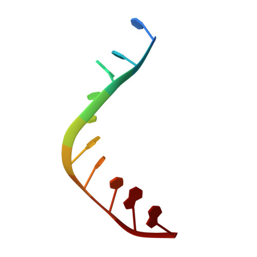 | ||
Sequence AnnotationsExpand | |||||
| |||||
| Ligands 2 Unique | |||||
|---|---|---|---|---|---|
| ID | Chains | Name / Formula / InChI Key | 2D Diagram | 3D Interactions | |
| KHN (Subject of Investigation/LOI) Query on KHN | D [auth A], F [auth B] | Ruthenium (bis-(phenanthroline)) (10-nitro-dipyridophenazine) C42 H26 N9 O2 Ru RRLMHIVRUDSXHL-UHFFFAOYSA-N |  | ||
| BA Query on BA | C [auth A], E [auth B] | BARIUM ION Ba XDFCIPNJCBUZJN-UHFFFAOYSA-N |  | ||
| Length ( Å ) | Angle ( ˚ ) |
|---|---|
| a = 46.73 | α = 90 |
| b = 46.73 | β = 90 |
| c = 31.99 | γ = 90 |
| Software Name | Purpose |
|---|---|
| PHENIX | refinement |
| xia2 | data reduction |
| xia2 | data scaling |
| PHASER | phasing |
| XDS | data reduction |
| XSCALE | data scaling |
| Coot | model building |
| Funding Organization | Location | Grant Number |
|---|---|---|
| Biotechnology and Biological Sciences Research Council | United Kingdom | BB/K019279/1 |
| Biotechnology and Biological Sciences Research Council | United Kingdom | BB/M004635/1 |
RCSB PDB (citation) is hosted by
RCSB PDB is a member of the

