6RMV

The crystal structure of a TRP channel peptide bound to a G protein beta gamma heterodimer


Experimental Data Snapshot

  • Method: X-RAY DIFFRACTION
  • Resolution: 1.94 Å
  • R-Value Free: 0.224 
  • R-Value Work: 0.188 
  • R-Value Observed: 0.190 

wwPDB Validation   3D Report Full Report


This is version 1.3 of the entry. See complete history


Literature

The structural basis for an on-off switch controlling G beta gamma-mediated inhibition of TRPM3 channels.

Behrendt, M.Gruss, F.Enzeroth, R.Dembla, S.Zhao, S.Crassous, P.A.Mohr, F.Nys, M.Louros, N.Gallardo, R.Zorzini, V.Wagner, D.Economou, A.Rousseau, F.Schymkowitz, J.Philipp, S.E.Rohacs, T.Ulens, C.Oberwinkler, J.

(2020) Proc Natl Acad Sci U S A 117: 29090-29100

  • DOI: https://doi.org/10.1073/pnas.2001177117
  • Primary Citation of Related Structures:  
    6RMV

  • PubMed Abstract: 

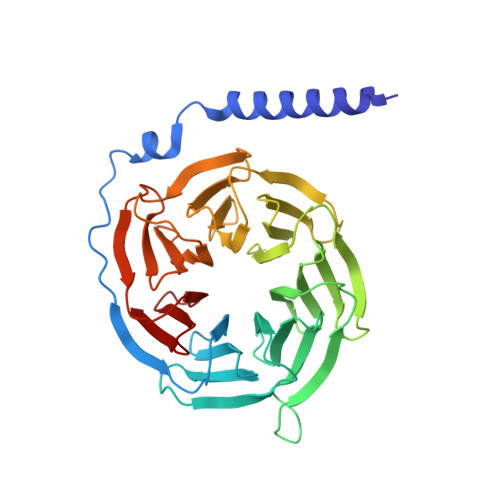
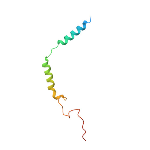
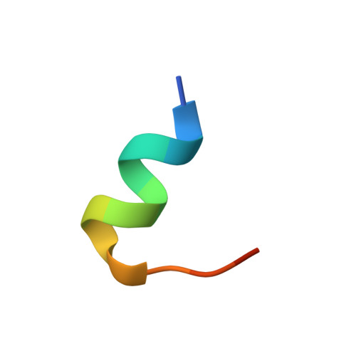
    TRPM3 channels play important roles in the detection of noxious heat and in inflammatory thermal hyperalgesia. The activity of these ion channels in somatosensory neurons is tightly regulated by µ-opioid receptors through the signaling of Gβγ proteins, thereby reducing TRPM3-mediated pain. We show here that Gβγ directly binds to a domain of 10 amino acids in TRPM3 and solve a cocrystal structure of this domain together with Gβγ. Using these data and mutational analysis of full-length proteins, we pinpoint three amino acids in TRPM3 and their interacting partners in Gβ 1 that are individually necessary for TRPM3 inhibition by Gβγ. The 10-amino-acid Gβγ-interacting domain in TRPM3 is subject to alternative splicing. Its inclusion in or exclusion from TRPM3 channel proteins therefore provides a mechanism for switching on or off the inhibitory action that Gβγ proteins exert on TRPM3 channels.


  • Organizational Affiliation

    Institut für Physiologie und Pathophysiologie, Philipps-Universität Marburg, 35037 Marburg, Germany.


Macromolecules
Find similar proteins by:  (by identity cutoff)  |  3D Structure
Entity ID: 1
MoleculeChains Sequence LengthOrganismDetailsImage
Guanine nucleotide-binding protein G(I)/G(S)/G(T) subunit beta-1340Mus musculusMutation(s): 0 
Gene Names: Gnb1
UniProt & NIH Common Fund Data Resources
Find proteins for P62874 (Mus musculus)
Explore P62874 
Go to UniProtKB:  P62874
IMPC:  MGI:95781
Entity Groups  
Sequence Clusters30% Identity50% Identity70% Identity90% Identity95% Identity100% Identity
UniProt GroupP62874
Sequence Annotations
Expand
  • Reference Sequence
Find similar proteins by:  (by identity cutoff)  |  3D Structure
Entity ID: 2
MoleculeChains Sequence LengthOrganismDetailsImage
Guanine nucleotide-binding protein G(I)/G(S)/G(O) subunit gamma-274Mus musculusMutation(s): 1 
Gene Names: Gng2
UniProt
Find proteins for P63213 (Mus musculus)
Explore P63213 
Go to UniProtKB:  P63213
Entity Groups  
Sequence Clusters30% Identity50% Identity70% Identity90% Identity95% Identity100% Identity
UniProt GroupP63213
Sequence Annotations
Expand
  • Reference Sequence

Find similar proteins by:  Sequence   |   3D Structure  

Entity ID: 3
MoleculeChains Sequence LengthOrganismDetailsImage
Transient receptor potential cation channel, subfamily M, member 315Mus musculusMutation(s): 0 
UniProt & NIH Common Fund Data Resources
Find proteins for Q2TV84 (Mus musculus)
Explore Q2TV84 
Go to UniProtKB:  Q2TV84
IMPC:  MGI:1330305
Entity Groups  
Sequence Clusters30% Identity50% Identity70% Identity90% Identity95% Identity100% Identity
UniProt GroupQ2TV84
Sequence Annotations
Expand
  • Reference Sequence
Small Molecules
Ligands 1 Unique
IDChains Name / Formula / InChI Key2D Diagram3D Interactions
GOL
Query on GOL

Download Ideal Coordinates CCD File 
D [auth B]GLYCEROL
C3 H8 O3
PEDCQBHIVMGVHV-UHFFFAOYSA-N
Experimental Data & Validation

Experimental Data

  • Method: X-RAY DIFFRACTION
  • Resolution: 1.94 Å
  • R-Value Free: 0.224 
  • R-Value Work: 0.188 
  • R-Value Observed: 0.190 
  • Space Group: P 21 21 21
Unit Cell:
Length ( Å )Angle ( ˚ )
a = 45.86α = 90
b = 80.432β = 90
c = 113.587γ = 90
Software Package:
Software NamePurpose
XDSdata reduction
Aimlessdata scaling
PHASERphasing
REFMACrefinement
PDB_EXTRACTdata extraction

Structure Validation

View Full Validation Report


Entry History & Funding Information

Deposition Data


Funding OrganizationLocationGrant Number
Fonds Wetenschappelijk Onderzoek - VlaanderenG0C9717N
European Union665501

Revision History  (Full details and data files)

  • Version 1.0: 2020-10-14
    Type: Initial release
  • Version 1.1: 2020-11-11
    Changes: Database references
  • Version 1.2: 2020-11-25
    Changes: Database references
  • Version 1.3: 2024-01-24
    Changes: Data collection, Database references, Refinement description