6RIQ

MinCD filament from Pseudomonas aeruginosa


Experimental Data Snapshot

  • Method: ELECTRON MICROSCOPY
  • Resolution: 3.10 Å
  • Aggregation State: HELICAL ARRAY 
  • Reconstruction Method: HELICAL 

wwPDB Validation   3D Report Full Report


This is version 1.1 of the entry. See complete history


Literature

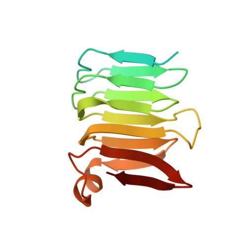
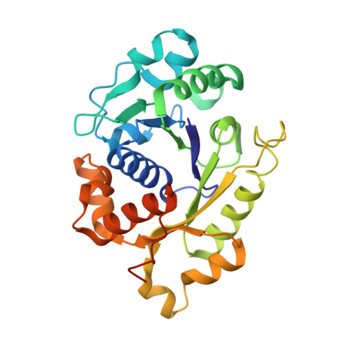
Cryo-EM structure of the MinCD copolymeric filament from Pseudomonas aeruginosa at 3.1 angstrom resolution.

Szewczak-Harris, A.Wagstaff, J.Lowe, J.

(2019) FEBS Lett 593: 1915-1926

  • DOI: https://doi.org/10.1002/1873-3468.13471
  • Primary Citation of Related Structures:  
    6RIQ

  • PubMed Abstract: 

    Positioning of the division site in many bacterial species relies on the MinCDE system, which prevents the cytokinetic Z-ring from assembling anywhere but the mid-cell, through an oscillatory diffusion-reaction mechanism. MinD dimers bind to membranes and, via their partner MinC, inhibit the polymerization of cell division protein FtsZ into the Z-ring. MinC and MinD form polymeric assemblies in solution and on cell membranes. Here, we report the high-resolution cryo-EM structure of the copolymeric filaments of Pseudomonas aeruginosa MinCD. The filaments consist of three protofilaments made of alternating MinC and MinD dimers. The MinCD protofilaments are almost completely straight and assemble as single protofilaments on lipid membranes, which we also visualized by cryo-EM.


  • Organizational Affiliation

    MRC Laboratory of Molecular Biology, Cambridge, UK.


Macromolecules
Find similar proteins by:  (by identity cutoff)  |  3D Structure
Entity ID: 1
MoleculeChains Sequence LengthOrganismDetailsImage
MinC
A, B, E, F, H
A, B, E, F, H, I, L, O, P, S, T
144Pseudomonas aeruginosaMutation(s): 0 
UniProt
Find proteins for Q9HYZ7 (Pseudomonas aeruginosa (strain ATCC 15692 / DSM 22644 / CIP 104116 / JCM 14847 / LMG 12228 / 1C / PRS 101 / PAO1))
Explore Q9HYZ7 
Go to UniProtKB:  Q9HYZ7
Entity Groups  
Sequence Clusters30% Identity50% Identity70% Identity90% Identity95% Identity100% Identity
UniProt GroupQ9HYZ7
Sequence Annotations
Expand
  • Reference Sequence
Find similar proteins by:  (by identity cutoff)  |  3D Structure
Entity ID: 2
MoleculeChains Sequence LengthOrganismDetailsImage
Site-determining protein
C, D, G, J, K
C, D, G, J, K, M, N, Q, R, U, V
271Pseudomonas aeruginosaMutation(s): 0 
Gene Names: 
UniProt
Find proteins for Q9HYZ6 (Pseudomonas aeruginosa (strain ATCC 15692 / DSM 22644 / CIP 104116 / JCM 14847 / LMG 12228 / 1C / PRS 101 / PAO1))
Explore Q9HYZ6 
Go to UniProtKB:  Q9HYZ6
Entity Groups  
Sequence Clusters30% Identity50% Identity70% Identity90% Identity95% Identity100% Identity
UniProt GroupQ9HYZ6
Sequence Annotations
Expand
  • Reference Sequence
Small Molecules
Ligands 2 Unique
IDChains Name / Formula / InChI Key2D Diagram3D Interactions
ATP
Query on ATP

Download Ideal Coordinates CCD File 
BA [auth G]
DA [auth J]
FA [auth K]
HA [auth M]
JA [auth N]
BA [auth G],
DA [auth J],
FA [auth K],
HA [auth M],
JA [auth N],
LA [auth Q],
NA [auth R],
PA [auth U],
RA [auth V],
X [auth C],
Z [auth D]
ADENOSINE-5'-TRIPHOSPHATE
C10 H16 N5 O13 P3
ZKHQWZAMYRWXGA-KQYNXXCUSA-N
MG
Query on MG

Download Ideal Coordinates CCD File 
AA [auth G]
CA [auth J]
EA [auth K]
GA [auth M]
IA [auth N]
AA [auth G],
CA [auth J],
EA [auth K],
GA [auth M],
IA [auth N],
KA [auth Q],
MA [auth R],
OA [auth U],
QA [auth V],
W [auth C],
Y [auth D]
MAGNESIUM ION
Mg
JLVVSXFLKOJNIY-UHFFFAOYSA-N
Experimental Data & Validation

Experimental Data

  • Method: ELECTRON MICROSCOPY
  • Resolution: 3.10 Å
  • Aggregation State: HELICAL ARRAY 
  • Reconstruction Method: HELICAL 

Structure Validation

View Full Validation Report


Entry History & Funding Information

Deposition Data


Funding OrganizationLocationGrant Number
Wellcome TrustUnited Kingdom202754/Z/16/Z
Medical Research Council (United Kingdom)United KingdomU105184326

Revision History  (Full details and data files)

  • Version 1.0: 2019-06-19
    Type: Initial release
  • Version 1.1: 2019-08-21
    Changes: Data collection, Database references