6RCY

CRYSTAL STRUCTURE OF FK1 DOMAIN OF FKBP52 IN COMPLEX WITH A BIO-INSPIRED HYBRID FLUORESCENT LIGAND


Experimental Data Snapshot

  • Method: X-RAY DIFFRACTION
  • Resolution: 2.30 Å
  • R-Value Free: 0.244 
  • R-Value Work: 0.218 
  • R-Value Observed: 0.220 

wwPDB Validation   3D Report Full Report


Ligand Structure Quality Assessment 


This is version 1.2 of the entry. See complete history


Literature

Bioinspired Hybrid Fluorescent Ligands for the FK1 Domain of FKBP52.

de la Sierra-Gallay, I.L.Belnou, M.Chambraud, B.Genet, M.van Tilbeurgh, H.Aumont-Nicaise, M.Desmadril, M.Baulieu, E.E.Jacquot, Y.Byrne, C.

(2020) J Med Chem 63: 10330-10338

  • DOI: https://doi.org/10.1021/acs.jmedchem.0c00825
  • Primary Citation of Related Structures:  
    6RCY

  • PubMed Abstract: 

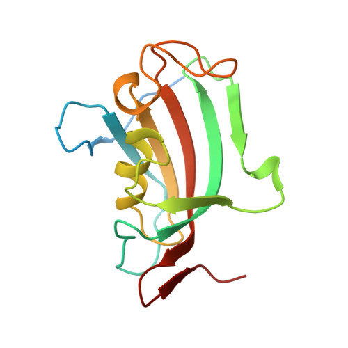
    The protein FKBP52 is a steroid hormone receptor coactivator likely involved in neurodegenerative disease. A series of small, water-soluble, bioinspired, pseudopeptidic fluorescent ligands for the FK1 domain of this protein are described. The design is such that engulfing of the ligand in the pocket of this domain is accompanied by hydrogen-bonding of the dansyl chromophore which functions as both an integral part of the ligand and a fluorescent reporter. Binding is concomitant with a significant wavelength shift and an enhancement of the ligand fluorescence signal. Excitation of FK1 domain native tryptophan residues in the presence of bound ligand results in Förster resonance energy transfer. Variation of key ligand residues within the short sequence was undertaken, and the interaction of the resulting library with the protein was measured by techniques including isothermal calorimetry analysis, fluorescence, and FRET quenching, and a range of K d values were determined. Cocrystallization of a protein ligand complex at 2.30 Å resolution provided detailed information at the atomic scale, while also providing insight into native substrate binding.


  • Organizational Affiliation

    Institut de Biologie Intégrative de la Cellule (I2BC), CNRS UMR9198, Université Paris-Saclay, Université Paris-Sud, 91405 Orsay, France.


Macromolecules
Find similar proteins by:  (by identity cutoff)  |  3D Structure
Entity ID: 1
MoleculeChains Sequence LengthOrganismDetailsImage
Peptidyl-prolyl cis-trans isomerase FKBP4148Homo sapiensMutation(s): 0 
Gene Names: FKBP4FKBP52
EC: 5.2.1.8
UniProt & NIH Common Fund Data Resources
Find proteins for Q02790 (Homo sapiens)
Explore Q02790 
Go to UniProtKB:  Q02790
PHAROS:  Q02790
GTEx:  ENSG00000004478 
Entity Groups  
Sequence Clusters30% Identity50% Identity70% Identity90% Identity95% Identity100% Identity
UniProt GroupQ02790
Sequence Annotations
Expand
  • Reference Sequence
Small Molecules
Ligands 1 Unique
IDChains Name / Formula / InChI Key2D Diagram3D Interactions
K0T
Query on K0T

Download Ideal Coordinates CCD File 
B [auth A](2~{S})-5-carbamimidamido-2-[[(2~{S})-2-[[(2~{S})-1-[5-(dimethylamino)naphthalen-1-yl]sulfonylpiperidin-2-yl]carbonylamino]-4-phenyl-butanoyl]amino]pentanoic acid
C34 H45 N7 O6 S
MKNCAFDCRIGOAN-YCVJPRETSA-N
Binding Affinity Annotations 
IDSourceBinding Affinity
K0T Binding MOAD:  6RCY Kd: 1600 (nM) from 1 assay(s)
Experimental Data & Validation

Experimental Data

  • Method: X-RAY DIFFRACTION
  • Resolution: 2.30 Å
  • R-Value Free: 0.244 
  • R-Value Work: 0.218 
  • R-Value Observed: 0.220 
  • Space Group: P 65 2 2
Unit Cell:
Length ( Å )Angle ( ˚ )
a = 52.96α = 90
b = 52.96β = 90
c = 237.86γ = 120
Software Package:
Software NamePurpose
BUSTERrefinement
XDSdata reduction
XDSdata scaling
PHASERphasing

Structure Validation

View Full Validation Report


Ligand Structure Quality Assessment 


Entry History 

Deposition Data

Revision History  (Full details and data files)

  • Version 1.0: 2020-05-13
    Type: Initial release
  • Version 1.1: 2020-10-21
    Changes: Database references
  • Version 1.2: 2024-01-24
    Changes: Data collection, Database references, Refinement description