6R9D

Crystal structure of an asymmetric dimer of the N-terminal domain of Euprosthenops australis Major Ampullate Spidroin 1 (dragline silk)


Experimental Data Snapshot

  • Method: X-RAY DIFFRACTION
  • Resolution: 2.10 Å
  • R-Value Free: 0.237 
  • R-Value Work: 0.208 
  • R-Value Observed: 0.209 

wwPDB Validation   3D Report Full Report


This is version 1.1 of the entry. See complete history


Literature

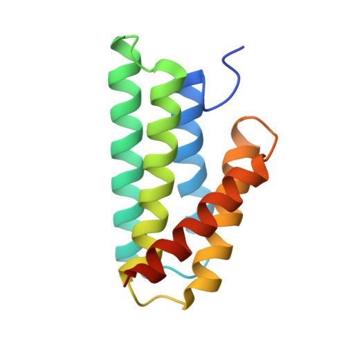
Structure of the N-terminal domain of Euprosthenops australis dragline silk suggests that conversion of spidroin dope to spider silk involves a conserved asymmetric dimer intermediate.

Jiang, W.Askarieh, G.Shkumatov, A.Hedhammar, M.Knight, S.D.

(2019) Acta Crystallogr D Struct Biol 75: 618-627

  • DOI: https://doi.org/10.1107/S2059798319007253
  • Primary Citation of Related Structures:  
    6R9D

  • PubMed Abstract: 

    Spider silk is a biomaterial with exceptional mechanical toughness, and there is great interest in developing biomimetic methods to produce engineered spider silk-based materials. However, the mechanisms that regulate the conversion of spider silk proteins (spidroins) from highly soluble dope into silk are not completely understood. The N-terminal domain (NT) of Euprosthenops australis dragline silk protein undergoes conformational and quaternary-structure changes from a monomer at a pH above 7 to a homodimer at lower pH values. Conversion from the monomer to the dimer requires the protonation of three conserved glutamic acid residues, resulting in a low-pH `locked' dimer stabilized by symmetric electrostatic interactions at the poles of the dimer. The detailed molecular events during this transition are still unresolved. Here, a 2.1 Å resolution crystal structure of an NT T61A mutant in an alternative, asymmetric, dimer form in which the electrostatic interactions at one of the poles are dramatically different from those in symmetrical dimers is presented. A similar asymmetric dimer structure from dragline silk of Nephila clavipes has previously been described. It is suggested that asymmetric dimers represent a conserved intermediate state in spider silk formation, and a revised `lock-and-trigger' mechanism for spider silk formation is presented.


  • Organizational Affiliation

    Department of Cell and Molecular Biology, Uppsala University, Biomedical Center, PO Box 596, SE-751 24 Uppsala, Sweden.


Macromolecules
Find similar proteins by:  (by identity cutoff)  |  3D Structure
Entity ID: 1
MoleculeChains Sequence LengthOrganismDetailsImage
Major ampullate spidroin 1
A, B, C, D, E
A, B, C, D, E, F
137Euprosthenops australisMutation(s): 1 
Gene Names: MaSp1
UniProt
Find proteins for Q05H60 (Euprosthenops australis)
Explore Q05H60 
Go to UniProtKB:  Q05H60
Entity Groups  
Sequence Clusters30% Identity50% Identity70% Identity90% Identity95% Identity100% Identity
UniProt GroupQ05H60
Sequence Annotations
Expand
  • Reference Sequence
Small Molecules
Ligands 1 Unique
IDChains Name / Formula / InChI Key2D Diagram3D Interactions
SO4
Query on SO4

Download Ideal Coordinates CCD File 
G [auth E]SULFATE ION
O4 S
QAOWNCQODCNURD-UHFFFAOYSA-L
Experimental Data & Validation

Experimental Data

  • Method: X-RAY DIFFRACTION
  • Resolution: 2.10 Å
  • R-Value Free: 0.237 
  • R-Value Work: 0.208 
  • R-Value Observed: 0.209 
  • Space Group: C 1 2 1
Unit Cell:
Length ( Å )Angle ( ˚ )
a = 126.631α = 90
b = 65.114β = 91.85
c = 95.968γ = 90
Software Package:
Software NamePurpose
PHENIXrefinement
XDSdata reduction
Aimlessdata scaling
PHENIXphasing

Structure Validation

View Full Validation Report


Entry History & Funding Information

Deposition Data


Funding OrganizationLocationGrant Number
Swedish Research CouncilSweden2016-04451

Revision History  (Full details and data files)

  • Version 1.0: 2019-07-17
    Type: Initial release
  • Version 1.1: 2024-01-24
    Changes: Data collection, Database references, Refinement description