6R0T

Getah virus macro domain in complex with ADPr in open conformation


Experimental Data Snapshot

  • Method: X-RAY DIFFRACTION
  • Resolution: 1.85 Å
  • R-Value Free: 0.199 
  • R-Value Work: 0.168 
  • R-Value Observed: 0.170 

wwPDB Validation   3D Report Full Report


Ligand Structure Quality Assessment 


This is version 1.3 of the entry. See complete history


Literature

Snapshots of ADP-ribose bound to Getah virus macro domain reveal an intriguing choreography.

Ferreira-Ramos, A.S.Sulzenbacher, G.Canard, B.Coutard, B.

(2020) Sci Rep 10: 14422-14422

  • DOI: https://doi.org/10.1038/s41598-020-70870-w
  • Primary Citation of Related Structures:  
    6QZU, 6R0F, 6R0G, 6R0P, 6R0R, 6R0T

  • PubMed Abstract: 

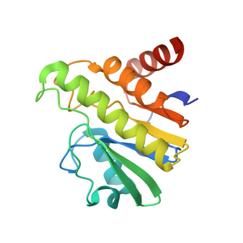
    Alphaviruses are (re-)emerging arboviruses of public health concern. The nsP3 gene product is one of the key players during viral replication. NsP3 comprises three domains: a macro domain, a zinc-binding domain and a hypervariable region. The macro domain is essential at both early and late stages of the replication cycle through ADP-ribose (ADPr) binding and de-ADP-ribosylation of host proteins. However, both its specific role and the precise molecular mechanism of de-ADP-ribosylation across specific viral families remains to be elucidated. Here we investigate by X-ray crystallography the mechanism of ADPr reactivity in the active site of Getah virus macro domain, which displays a peculiar substitution of one of the conserved residues in the catalytic loop. ADPr adopts distinct poses including a covalent bond between the C''1 of the ADPr and a conserved Togaviridae-specific cysteine. These different poses observed for ADPr may represent snapshots of the de-ADP-ribosylation mechanism, highlighting residues to be further characterised.


  • Organizational Affiliation

    Architecture et Fonction des Macromolécules Biologiques, CNRS, Aix-Marseille Université, 13288, Marseille, France.


Macromolecules
Find similar proteins by:  (by identity cutoff)  |  3D Structure
Entity ID: 1
MoleculeChains Sequence LengthOrganismDetailsImage
Non-structural polyprotein
A, B
168Getah virusMutation(s): 0 
Gene Names: nsP1234
UniProt
Find proteins for Q5Y389 (Getah virus)
Explore Q5Y389 
Go to UniProtKB:  Q5Y389
Entity Groups  
Sequence Clusters30% Identity50% Identity70% Identity90% Identity95% Identity100% Identity
UniProt GroupQ5Y389
Sequence Annotations
Expand
  • Reference Sequence
Small Molecules
Ligands 3 Unique
IDChains Name / Formula / InChI Key2D Diagram3D Interactions
JNT
Query on JNT

Download Ideal Coordinates CCD File 
C [auth A],
I [auth B]
[[(2~{R},3~{S},4~{R},5~{R})-5-(6-aminopurin-9-yl)-3,4-bis(oxidanyl)oxolan-2-yl]methoxy-oxidanyl-phosphoryl] [(2~{R},3~{S},4~{S})-2,3,4,5-tetrakis(oxidanyl)pentyl] hydrogen phosphate
C15 H25 N5 O14 P2
QKJRZYRQRSLXSV-AOOZFPJJSA-N
EDO
Query on EDO

Download Ideal Coordinates CCD File 
D [auth A],
E [auth A],
F [auth A],
J [auth B]
1,2-ETHANEDIOL
C2 H6 O2
LYCAIKOWRPUZTN-UHFFFAOYSA-N
ACT
Query on ACT

Download Ideal Coordinates CCD File 
G [auth A],
H [auth A]
ACETATE ION
C2 H3 O2
QTBSBXVTEAMEQO-UHFFFAOYSA-M
Experimental Data & Validation

Experimental Data

  • Method: X-RAY DIFFRACTION
  • Resolution: 1.85 Å
  • R-Value Free: 0.199 
  • R-Value Work: 0.168 
  • R-Value Observed: 0.170 
  • Space Group: P 21 21 21
Unit Cell:
Length ( Å )Angle ( ˚ )
a = 46.673α = 90
b = 71.441β = 90
c = 98.97γ = 90
Software Package:
Software NamePurpose
REFMACrefinement
XDSdata reduction
Aimlessdata scaling
REFMACphasing

Structure Validation

View Full Validation Report


Ligand Structure Quality Assessment 


Entry History 

Deposition Data

Revision History  (Full details and data files)

  • Version 1.0: 2020-04-01
    Type: Initial release
  • Version 1.1: 2020-10-14
    Changes: Database references
  • Version 1.2: 2022-12-21
    Changes: Data collection, Database references
  • Version 1.3: 2024-01-31
    Changes: Data collection, Refinement description