6QFY

CRYSTAL STRUCTURE OF PORCINE HEMAGGLUTINATING ENCEPHALOMYELITIS VIRUS SPIKE PROTEIN LECTIN DOMAIN


Experimental Data Snapshot

  • Method: X-RAY DIFFRACTION
  • Resolution: 2.97 Å
  • R-Value Free: 0.255 
  • R-Value Work: 0.218 
  • R-Value Observed: 0.220 

wwPDB Validation   3D Report Full Report


Ligand Structure Quality Assessment 


This is version 2.1 of the entry. See complete history


Literature

Human coronaviruses OC43 and HKU1 bind to 9-O-acetylated sialic acids via a conserved receptor-binding site in spike protein domain A.

Hulswit, R.J.G.Lang, Y.Bakkers, M.J.G.Li, W.Li, Z.Schouten, A.Ophorst, B.van Kuppeveld, F.J.M.Boons, G.J.Bosch, B.J.Huizinga, E.G.de Groot, R.J.

(2019) Proc Natl Acad Sci U S A 116: 2681-2690

  • DOI: https://doi.org/10.1073/pnas.1809667116
  • Primary Citation of Related Structures:  
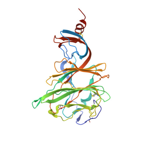
    6QFY

  • PubMed Abstract: 

    Human betacoronaviruses OC43 and HKU1 are endemic respiratory pathogens and, while related, originated from independent zoonotic introductions. OC43 is in fact a host-range variant of the species Betacoronavirus-1 , and more closely related to bovine coronavirus (BCoV)-its presumptive ancestor-and porcine hemagglutinating encephalomyelitis virus (PHEV). The β1-coronaviruses (β1CoVs) and HKU1 employ glycan-based receptors carrying 9- O -acetylated sialic acid (9- O -Ac-Sia). Receptor binding is mediated by spike protein S, the main determinant of coronavirus host specificity. For BCoV, a crystal structure for the receptor-binding domain S1 A is available and for HKU1 a cryoelectron microscopy structure of the complete S ectodomain. However, the location of the receptor-binding site (RBS), arguably the single-most important piece of information, is unknown. Here we solved the 3.0-Å crystal structure of PHEV S1 A We then took a comparative structural analysis approach to map the β1CoV S RBS, using the general design of 9- O -Ac-Sia-binding sites as blueprint, backed-up by automated ligand docking, structure-guided mutagenesis of OC43, BCoV, and PHEV S1 A , and infectivity assays with BCoV-S-pseudotyped vesicular stomatitis viruses. The RBS is not exclusive to OC43 and related animal viruses, but is apparently conserved and functional also in HKU1 S1 A The binding affinity of the HKU1 S RBS toward short sialoglycans is significantly lower than that of OC43, which we attribute to differences in local architecture and accessibility, and which may be indicative for differences between the two viruses in receptor fine-specificity. Our findings challenge reports that would map the OC43 RBS elsewhere in S1 A and that of HKU1 in domain S1 B .


  • Organizational Affiliation

    Virology Division, Department of Infectious Diseases and Immunology, Faculty of Veterinary Medicine, Utrecht University, 3584 CH Utrecht, The Netherlands.


Macromolecules
Find similar proteins by:  (by identity cutoff)  |  3D Structure
Entity ID: 1
MoleculeChains Sequence LengthOrganismDetailsImage
Spike glycoprotein
A, B
288Porcine hemagglutinating encephalomyelitis virusMutation(s): 0 
UniProt
Find proteins for A0A1Z2WUW0 (Porcine hemagglutinating encephalomyelitis virus)
Explore A0A1Z2WUW0 
Go to UniProtKB:  A0A1Z2WUW0
Entity Groups  
Sequence Clusters30% Identity50% Identity70% Identity90% Identity95% Identity100% Identity
UniProt GroupA0A1Z2WUW0
Sequence Annotations
Expand
  • Reference Sequence
Oligosaccharides

Help

Entity ID: 2
MoleculeChains Length2D Diagram Glycosylation3D Interactions
2-acetamido-2-deoxy-beta-D-glucopyranose-(1-4)-2-acetamido-2-deoxy-beta-D-glucopyranose
C, D, F, G
2N-Glycosylation
Glycosylation Resources
GlyTouCan:  G42666HT
GlyCosmos:  G42666HT
GlyGen:  G42666HT
Entity ID: 3
MoleculeChains Length2D Diagram Glycosylation3D Interactions
beta-D-mannopyranose-(1-4)-2-acetamido-2-deoxy-beta-D-glucopyranose-(1-4)-2-acetamido-2-deoxy-beta-D-glucopyranose
E
3N-Glycosylation
Glycosylation Resources
GlyTouCan:  G15407YE
GlyCosmos:  G15407YE
GlyGen:  G15407YE
Experimental Data & Validation

Experimental Data

  • Method: X-RAY DIFFRACTION
  • Resolution: 2.97 Å
  • R-Value Free: 0.255 
  • R-Value Work: 0.218 
  • R-Value Observed: 0.220 
  • Space Group: P 31 2 1
Unit Cell:
Length ( Å )Angle ( ˚ )
a = 112.572α = 90
b = 112.572β = 90
c = 141.441γ = 120
Software Package:
Software NamePurpose
REFMACrefinement
PDB_EXTRACTdata extraction
XDSdata reduction
Aimlessdata scaling
PHASERphasing

Structure Validation

View Full Validation Report


Ligand Structure Quality Assessment 


Entry History 

Deposition Data

Revision History  (Full details and data files)

  • Version 1.0: 2019-02-06
    Type: Initial release
  • Version 1.1: 2019-02-20
    Changes: Data collection, Database references
  • Version 2.0: 2020-07-29
    Type: Remediation
    Reason: Carbohydrate remediation
    Changes: Atomic model, Data collection, Derived calculations, Structure summary
  • Version 2.1: 2024-01-24
    Changes: Data collection, Database references, Refinement description, Structure summary