6P73

Cytochrome-C-nitrite reductase


Experimental Data Snapshot

  • Method: X-RAY DIFFRACTION
  • Resolution: 1.65 Å
  • R-Value Free: 0.211 
  • R-Value Work: 0.171 
  • R-Value Observed: 0.172 

wwPDB Validation   3D Report Full Report

Currently 6P73 does not have a validation slider image.


This is version 1.1 of the entry. See complete history


Literature

Trapping of a Putative Intermediate in the CytochromecNitrite Reductase (ccNiR)-Catalyzed Reduction of Nitrite: Implications for the ccNiR Reaction Mechanism.

Ali, M.Stein, N.Mao, Y.Shahid, S.Schmidt, M.Bennett, B.Pacheco, A.A.

(2019) J Am Chem Soc 141: 13358-13371

  • DOI: https://doi.org/10.1021/jacs.9b03036
  • Primary Citation of Related Structures:  
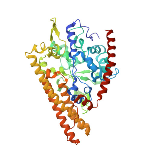
    6P73

  • PubMed Abstract: 

    Cytochrome c nitrite reductase (ccNiR) is a periplasmic, decaheme homodimeric enzyme that catalyzes the six-electron reduction of nitrite to ammonia. Under standard assay conditions catalysis proceeds without detected intermediates, and it has been assumed that this is also true in vivo. However, this report demonstrates that it is possible to trap a putative intermediate by controlling the electrochemical potential at which reduction takes place. UV/vis spectropotentiometry showed that nitrite-loaded Shewanella oneidensis ccNiR is reduced in a concerted two-electron step to generate an {FeNO} 7 moiety at the active site, with an associated midpoint potential of +246 mV vs SHE at pH 7. By contrast, cyanide-bound active site reduction is a one-electron process with a midpoint potential of +20 mV, and without a strong-field ligand the active site midpoint potential shifts 70 mV lower still. EPR analysis subsequently revealed that the {FeNO} 7 moiety possesses an unusual spectral signature, different from those normally observed for {FeNO} 7 hemes, that may indicate magnetic interaction of the active site with nearby hemes. Protein film voltammetry experiments previously showed that catalytic nitrite reduction to ammonia by S. oneidensis ccNiR requires an applied potential of at least -120 mV, well below the midpoint potential for {FeNO} 7 formation. Thus, it appears that an {FeNO} 7 active site is a catalytic intermediate in the ccNiR-mediated reduction of nitrite to ammonia, whose degree of accumulation depends exclusively on the applied potential. At low potentials the species is rapidly reduced and does not accumulate, while at higher potentials it is trapped, thus preventing catalytic ammonia formation.


  • Organizational Affiliation

    Department of Chemistry and Biochemistry , University of Wisconsin-Milwaukee , Milwaukee , Wisconsin 53211 , United States.


Macromolecules
Find similar proteins by:  (by identity cutoff)  |  3D Structure
Entity ID: 1
MoleculeChains Sequence LengthOrganismDetailsImage
Cytochrome c-552
A, B
439Shewanella oneidensisMutation(s): 0 
Gene Names: nrfASO_3980
EC: 1.7.2.2
UniProt
Find proteins for Q8EAC7 (Shewanella oneidensis (strain MR-1))
Explore Q8EAC7 
Go to UniProtKB:  Q8EAC7
Entity Groups  
Sequence Clusters30% Identity50% Identity70% Identity90% Identity95% Identity100% Identity
UniProt GroupQ8EAC7
Sequence Annotations
Expand
  • Reference Sequence
Small Molecules
Ligands 2 Unique
IDChains Name / Formula / InChI Key2D Diagram3D Interactions
HEC (Subject of Investigation/LOI)
Query on HEC

Download Ideal Coordinates CCD File 
C [auth A]
D [auth A]
E [auth A]
F [auth A]
G [auth A]
C [auth A],
D [auth A],
E [auth A],
F [auth A],
G [auth A],
I [auth B],
J [auth B],
K [auth B],
L [auth B],
M [auth B]
HEME C
C34 H34 Fe N4 O4
HXQIYSLZKNYNMH-LJNAALQVSA-N
CA
Query on CA

Download Ideal Coordinates CCD File 
H [auth A],
N [auth B]
CALCIUM ION
Ca
BHPQYMZQTOCNFJ-UHFFFAOYSA-N
Experimental Data & Validation

Experimental Data

  • Method: X-RAY DIFFRACTION
  • Resolution: 1.65 Å
  • R-Value Free: 0.211 
  • R-Value Work: 0.171 
  • R-Value Observed: 0.172 
  • Space Group: P 21 21 21
Unit Cell:
Length ( Å )Angle ( ˚ )
a = 50.18α = 90
b = 96.07β = 90
c = 222.6γ = 90
Software Package:
Software NamePurpose
PHENIXrefinement
MOSFLMdata reduction
SCALAdata scaling
PHENIXphasing

Structure Validation

View Full Validation Report

Currently 6P73 does not have a validation slider image.


Entry History & Funding Information

Deposition Data


Funding OrganizationLocationGrant Number
National Science Foundation (NSF, United States)United States--

Revision History  (Full details and data files)

  • Version 1.0: 2020-04-01
    Type: Initial release
  • Version 1.1: 2023-10-11
    Changes: Data collection, Database references, Derived calculations, Refinement description