6OV8

2.6 Angstrom Resolution Crystal Structure of Aminopeptidase B from Escherichia coli str. K-12 substr. MG1655


Experimental Data Snapshot

  • Method: X-RAY DIFFRACTION
  • Resolution: 2.61 Å
  • R-Value Free: 0.238 
  • R-Value Work: 0.181 
  • R-Value Observed: 0.184 

wwPDB Validation   3D Report Full Report


This is version 1.4 of the entry. See complete history


Literature

Comparison of metal-bound and unbound structures of aminopeptidase B proteins from Escherichia coli and Yersinia pestis.

Minasov, G.Lam, M.R.Rosas-Lemus, M.Slawek, J.Woinska, M.Shabalin, I.G.Shuvalova, L.Palsson, B.O.Godzik, A.Minor, W.Satchell, K.J.F.

(2020) Protein Sci 29: 1618-1628

  • DOI: https://doi.org/10.1002/pro.3876
  • Primary Citation of Related Structures:  
    6OAD, 6OV8

  • PubMed Abstract: 

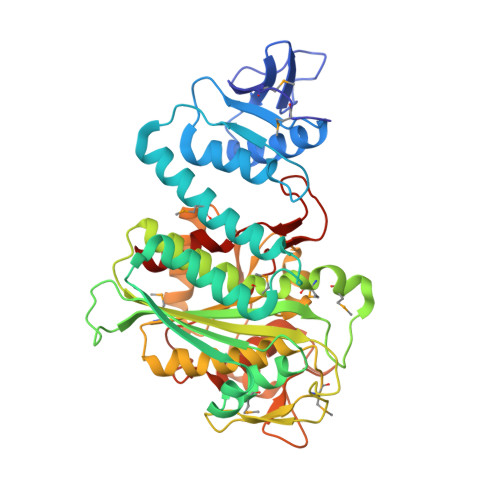
    Protein degradation by aminopeptidases is involved in bacterial responses to stress. Escherichia coli produces two metal-dependent M17 family leucine aminopeptidases (LAPs), aminopeptidase A (PepA) and aminopeptidase B (PepB). Several structures have been solved for PepA as well as other bacterial M17 peptidases. Herein, we report the first structures of a PepB M17 peptidase. The E. coli PepB protein structure was determined at a resolution of 2.05 and 2.6 Å. One structure has both Zn 2+ and Mn 2+ , while the second structure has two Zn 2+ ions bound to the active site. A 2.75 Å apo structure is also reported for PepB from Yersinia pestis. Both proteins form homohexamers, similar to the overall arrangement of PepA and other M17 peptidases. However, the divergent N-terminal domain in PepB is much larger resulting in a tertiary structure that is more expanded. Modeling of a dipeptide substrate into the C-terminal LAP domain reveals contacts that account for PepB to uniquely cleave after aspartate.


  • Organizational Affiliation

    Department of Microbiology-Immunology, Feinberg School of Medicine, Northwestern University, Chicago, Illinois, USA.


Macromolecules
Find similar proteins by:  (by identity cutoff)  |  3D Structure
Entity ID: 1
MoleculeChains Sequence LengthOrganismDetailsImage
Peptidase B
A, B, C, D, E
A, B, C, D, E, F
430Escherichia coli K-12Mutation(s): 0 
Gene Names: pepByfhIb2523JW2507
EC: 3.4.11.23
UniProt
Find proteins for P37095 (Escherichia coli (strain K12))
Explore P37095 
Go to UniProtKB:  P37095
Entity Groups  
Sequence Clusters30% Identity50% Identity70% Identity90% Identity95% Identity100% Identity
UniProt GroupP37095
Sequence Annotations
Expand
  • Reference Sequence
Small Molecules
Ligands 3 Unique
IDChains Name / Formula / InChI Key2D Diagram3D Interactions
ZN
Query on ZN

Download Ideal Coordinates CCD File 
CA [auth E]
G [auth A]
HA [auth F]
M [auth B]
S [auth C]
CA [auth E],
G [auth A],
HA [auth F],
M [auth B],
S [auth C],
X [auth D]
ZINC ION
Zn
PTFCDOFLOPIGGS-UHFFFAOYSA-N
MN (Subject of Investigation/LOI)
Query on MN

Download Ideal Coordinates CCD File 
DA [auth E]
H [auth A]
IA [auth F]
N [auth B]
T [auth C]
DA [auth E],
H [auth A],
IA [auth F],
N [auth B],
T [auth C],
Y [auth D]
MANGANESE (II) ION
Mn
WAEMQWOKJMHJLA-UHFFFAOYSA-N
CL
Query on CL

Download Ideal Coordinates CCD File 
AA [auth D]
BA [auth D]
EA [auth E]
FA [auth E]
GA [auth E]
AA [auth D],
BA [auth D],
EA [auth E],
FA [auth E],
GA [auth E],
I [auth A],
J [auth A],
JA [auth F],
K [auth A],
KA [auth F],
L [auth A],
LA [auth F],
MA [auth F],
O [auth B],
P [auth B],
Q [auth B],
R [auth B],
U [auth C],
V [auth C],
W [auth C],
Z [auth D]
CHLORIDE ION
Cl
VEXZGXHMUGYJMC-UHFFFAOYSA-M
Modified Residues  1 Unique
IDChains TypeFormula2D DiagramParent
MSE
Query on MSE
A, B, C, D, E
A, B, C, D, E, F
L-PEPTIDE LINKINGC5 H11 N O2 SeMET
Experimental Data & Validation

Experimental Data

  • Method: X-RAY DIFFRACTION
  • Resolution: 2.61 Å
  • R-Value Free: 0.238 
  • R-Value Work: 0.181 
  • R-Value Observed: 0.184 
  • Space Group: P 21 21 21
Unit Cell:
Length ( Å )Angle ( ˚ )
a = 114.857α = 90
b = 148.19β = 90
c = 165.01γ = 90
Software Package:
Software NamePurpose
REFMACrefinement
HKL-3000data reduction
HKL-3000data scaling
PHASERphasing

Structure Validation

View Full Validation Report


Entry History 

Revision History  (Full details and data files)

  • Version 1.0: 2019-05-15
    Type: Initial release
  • Version 1.1: 2021-01-27
    Changes: Database references, Derived calculations
  • Version 1.2: 2021-04-07
    Changes: Database references
  • Version 1.3: 2023-10-11
    Changes: Data collection, Database references, Refinement description
  • Version 1.4: 2023-11-15
    Changes: Data collection