Cryo-EM structure of OSCA1.2 fromOryza sativaelucidates the mechanical basis of potential membrane hyperosmolality gating.
Maity, K., Heumann, J.M., McGrath, A.P., Kopcho, N.J., Hsu, P.K., Lee, C.W., Mapes, J.H., Garza, D., Krishnan, S., Morgan, G.P., Hendargo, K.J., Klose, T., Rees, S.D., Medrano-Soto, A., Saier Jr., M.H., Pineros, M., Komives, E.A., Schroeder, J.I., Chang, G., Stowell, M.H.B.(2019) Proc Natl Acad Sci U S A 116: 14309-14318
- PubMed: 31227607
- DOI: https://doi.org/10.1073/pnas.1900774116
- Primary Citation of Related Structures:
6OCE - PubMed Abstract:
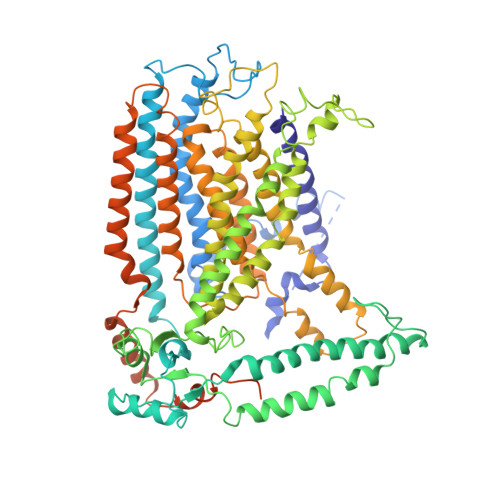
Sensing and responding to environmental water deficiency and osmotic stresses are essential for the growth, development, and survival of plants. Recently, an osmolality-sensing ion channel called OSCA1 was discovered that functions in sensing hyperosmolality in Arabidopsis Here, we report the cryo-electron microscopy (cryo-EM) structure and function of an OSCA1 homolog from rice ( Oryza sativa ; OsOSCA1.2), leading to a model of how it could mediate hyperosmolality sensing and transport pathway gating. The structure reveals a dimer; the molecular architecture of each subunit consists of 11 transmembrane (TM) helices and a cytosolic soluble domain that has homology to RNA recognition proteins. The TM domain is structurally related to the TMEM16 family of calcium-dependent ion channels and lipid scramblases. The cytosolic soluble domain possesses a distinct structural feature in the form of extended intracellular helical arms that are parallel to the plasma membrane. These helical arms are well positioned to potentially sense lateral tension on the inner leaflet of the lipid bilayer caused by changes in turgor pressure. Computational dynamic analysis suggests how this domain couples to the TM portion of the molecule to open a transport pathway. Hydrogen/deuterium exchange mass spectrometry (HDXMS) experimentally confirms the conformational dynamics of these coupled domains. These studies provide a framework to understand the structural basis of proposed hyperosmolality sensing in a staple crop plant, extend our knowledge of the anoctamin superfamily important for plants and fungi, and provide a structural mechanism for potentially translating membrane stress to transport regulation.
Organizational Affiliation:
Skaggs School of Pharmacy and Pharmaceutical Sciences, University of California San Diego, La Jolla, CA 92093.