6O3H

Icosahedral reconstruction of the thermophilic bacteriophage P74-26 capsid


Experimental Data Snapshot

  • Method: ELECTRON MICROSCOPY
  • Resolution: 2.80 Å
  • Aggregation State: PARTICLE 
  • Reconstruction Method: SINGLE PARTICLE 

wwPDB Validation   3D Report Full Report


This is version 1.3 of the entry. See complete history


Literature

Principles for enhancing virus capsid capacity and stability from a thermophilic virus capsid structure.

Stone, N.P.Demo, G.Agnello, E.Kelch, B.A.

(2019) Nat Commun 10: 4471-4471

  • DOI: https://doi.org/10.1038/s41467-019-12341-z
  • Primary Citation of Related Structures:  
    6O3H

  • PubMed Abstract: 

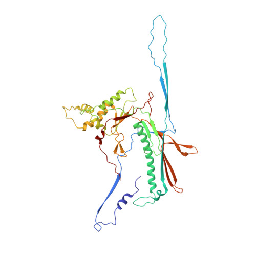
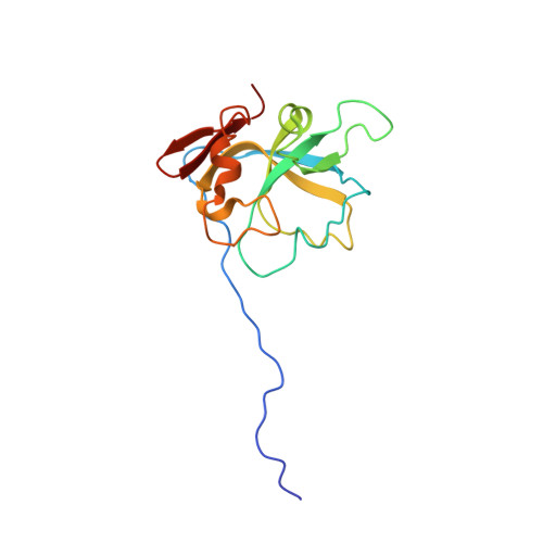
    The capsids of double-stranded DNA viruses protect the viral genome from the harsh extracellular environment, while maintaining stability against the high internal pressure of packaged DNA. To elucidate how capsids maintain stability in an extreme environment, we use cryoelectron microscopy to determine the capsid structure of thermostable phage P74-26 to 2.8-Å resolution. We find P74-26 capsids exhibit an overall architecture very similar to those of other tailed bacteriophages, allowing us to directly compare structures to derive the structural basis for enhanced stability. Our structure reveals lasso-like interactions that appear to function like catch bonds. This architecture allows the capsid to expand during genome packaging, yet maintain structural stability. The P74-26 capsid has T = 7 geometry despite being twice as large as mesophilic homologs. Capsid capacity is increased with a larger, flatter major capsid protein. Given these results, we predict decreased icosahedral complexity (i.e. T ≤ 7) leads to a more stable capsid assembly.


  • Organizational Affiliation

    Department of Biochemistry and Molecular Pharmacology, University of Massachusetts Medical School, Worcester, MA, 01655, USA.


Macromolecules
Find similar proteins by:  (by identity cutoff)  |  3D Structure
Entity ID: 1
MoleculeChains Sequence LengthOrganismDetailsImage
Major head protein
A, B, C, D, E
A, B, C, D, E, F, G
409Oshimavirus P7426Mutation(s): 0 
UniProt
Find proteins for A7XXR6 (Thermus virus P74-26)
Explore A7XXR6 
Go to UniProtKB:  A7XXR6
Entity Groups  
Sequence Clusters30% Identity50% Identity70% Identity90% Identity95% Identity100% Identity
UniProt GroupA7XXR6
Sequence Annotations
Expand
  • Reference Sequence
Find similar proteins by:  (by identity cutoff)  |  3D Structure
Entity ID: 2
MoleculeChains Sequence LengthOrganismDetailsImage
P74-26 Head Decoration Protein
H, I, J, K, L
H, I, J, K, L, M, N
146Oshimavirus P7426Mutation(s): 0 
UniProt
Find proteins for A7XXR5 (Thermus virus P74-26)
Explore A7XXR5 
Go to UniProtKB:  A7XXR5
Entity Groups  
Sequence Clusters30% Identity50% Identity70% Identity90% Identity95% Identity100% Identity
UniProt GroupA7XXR5
Sequence Annotations
Expand
  • Reference Sequence
Experimental Data & Validation

Experimental Data

  • Method: ELECTRON MICROSCOPY
  • Resolution: 2.80 Å
  • Aggregation State: PARTICLE 
  • Reconstruction Method: SINGLE PARTICLE 
EM Software:
TaskSoftware PackageVersion
RECONSTRUCTIONFREALIGN9.11
MODEL REFINEMENTPHENIX1.14

Structure Validation

View Full Validation Report


Entry History & Funding Information

Deposition Data


Funding OrganizationLocationGrant Number
National Science Foundation (NSF, United States)United States--

Revision History  (Full details and data files)

  • Version 1.0: 2019-07-24
    Type: Initial release
  • Version 1.1: 2019-10-23
    Changes: Data collection, Database references
  • Version 1.2: 2019-11-27
    Changes: Author supporting evidence
  • Version 1.3: 2024-03-20
    Changes: Data collection, Database references, Derived calculations