6N9O

Crystal structure of human GSDMD


Experimental Data Snapshot

  • Method: X-RAY DIFFRACTION
  • Resolution: 3.50 Å
  • R-Value Free: 0.345 
  • R-Value Work: 0.276 
  • R-Value Observed: 0.280 

wwPDB Validation   3D Report Full Report

Currently 6N9O does not have a validation slider image.


This is version 1.3 of the entry. See complete history


Literature

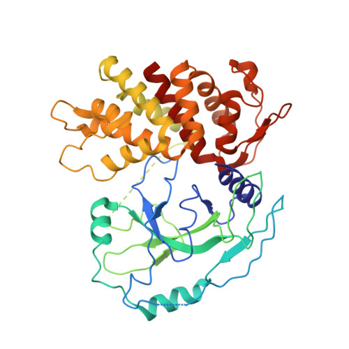
Crystal Structures of the Full-Length Murine and Human Gasdermin D Reveal Mechanisms of Autoinhibition, Lipid Binding, and Oligomerization.

Liu, Z.Wang, C.Yang, J.Zhou, B.Yang, R.Ramachandran, R.Abbott, D.W.Xiao, T.S.

(2019) Immunity 51: 43

  • DOI: https://doi.org/10.1016/j.immuni.2019.04.017
  • Primary Citation of Related Structures:  
    6N9N, 6N9O

  • PubMed Abstract: 

    Gasdermin D (GSDMD) is an effector molecule for pyroptosis downstream of canonical and noncanonical inflammasome signaling pathways. Cleavage of GSDMD by inflammatory caspases triggers the oligomerization and lipid binding by its N-terminal domain, which assembles membrane pores, whereas its C-terminal domain binds the N-terminal domain to inhibit pyroptosis. Despite recent progress in our understanding of the structure and function of the murine gasdermin A3 (mGSDMA3), the molecular mechanisms of GSDMD activation and regulation remain poorly characterized. Here, we report the crystal structures of the full-length murine and human GSDMDs, which reveal the architecture of the GSDMD N-terminal domains and demonstrate distinct and common features of autoinhibition among gasdermin family members utilizing their β1-β2 loops. Disruption of the intramolecular domain interface enhanced pyroptosis, whereas mutations at the predicted lipid-binding or oligomerization surface reduced cytolysis. Our study provides a framework for understanding the autoinhibition, lipid binding, and oligomerization of GSDMD by using overlapping interfaces.


  • Organizational Affiliation

    Department of Pathology, Case Western Reserve University, Cleveland, OH 44106 USA.


Macromolecules
Find similar proteins by:  (by identity cutoff)  |  3D Structure
Entity ID: 1
MoleculeChains Sequence LengthOrganismDetailsImage
Gasdermin-D
A, B, C, D
485Homo sapiensMutation(s): 0 
Gene Names: GSDMDDFNA5LGSDMDC1FKSG10
UniProt & NIH Common Fund Data Resources
Find proteins for P57764 (Homo sapiens)
Explore P57764 
Go to UniProtKB:  P57764
PHAROS:  P57764
GTEx:  ENSG00000104518 
Entity Groups  
Sequence Clusters30% Identity50% Identity70% Identity90% Identity95% Identity100% Identity
UniProt GroupP57764
Sequence Annotations
Expand
  • Reference Sequence
Experimental Data & Validation

Experimental Data

  • Method: X-RAY DIFFRACTION
  • Resolution: 3.50 Å
  • R-Value Free: 0.345 
  • R-Value Work: 0.276 
  • R-Value Observed: 0.280 
  • Space Group: P 1 21 1
Unit Cell:
Length ( Å )Angle ( ˚ )
a = 58.64α = 90
b = 105.55β = 94.47
c = 151.79γ = 90
Software Package:
Software NamePurpose
PHENIXrefinement
XDSdata reduction
XSCALEdata scaling
PHENIXphasing

Structure Validation

View Full Validation Report

Currently 6N9O does not have a validation slider image.


Entry History & Funding Information

Deposition Data


Funding OrganizationLocationGrant Number
National Institutes of Health/National Institute of Arthritis and Musculoskeletal and Skin Diseases (NIH/NIAMS)United StatesGM127609
National Institutes of Health/National Institute of Arthritis and Musculoskeletal and Skin Diseases (NIH/NIAMS)United StatesAR069908

Revision History  (Full details and data files)

  • Version 1.0: 2019-06-05
    Type: Initial release
  • Version 1.1: 2019-07-31
    Changes: Data collection, Database references
  • Version 1.2: 2019-12-11
    Changes: Author supporting evidence
  • Version 1.3: 2023-10-11
    Changes: Data collection, Database references, Refinement description