6MHM

Crystal structure of human acid ceramidase in covalent complex with carmofur


Experimental Data Snapshot

  • Method: X-RAY DIFFRACTION
  • Resolution: 2.74 Å
  • R-Value Free: 0.262 
  • R-Value Work: 0.214 
  • R-Value Observed: 0.218 

wwPDB Validation   3D Report Full Report

Currently 6MHM does not have a validation slider image.


This is version 2.1 of the entry. See complete history


Literature

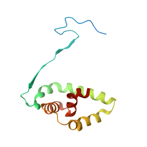
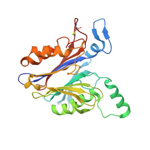
Molecular Mechanism of Inhibition of Acid Ceramidase by Carmofur.

Dementiev, A.Joachimiak, A.Nguyen, H.Gorelik, A.Illes, K.Shabani, S.Gelsomino, M.Ahn, E.E.Nagar, B.Doan, N.

(2019) J Med Chem 62: 987-992

  • DOI: https://doi.org/10.1021/acs.jmedchem.8b01723
  • Primary Citation of Related Structures:  
    6MHM

  • PubMed Abstract: 

    Human acid ceramidase (AC) is a lysosomal cysteine amidase, which has received a great deal of interest in recent years as a potential target for the development of new therapeutics against melanoma and glioblastoma tumors. Despite the strong interest in obtaining structural information, only the structures of the apo-AC enzyme in its zymogen and activated conformations are available. In this work, the crystal structure of AC in complex with the covalent carmofur inhibitor is presented. Carmofur is an antineoplastic drug containing an electrophilic carbonyl reactive group that targets the catalytic cysteine. This novel structural data explains the basis of the AC inhibition, provides insights into the enzymatic properties of the protein, and is a great aid toward the structure-based drug design of potent inhibitors for AC, providing the detailed mechanism, which has eluded the scientific community for more than 30 years, of carmofur's mysterious 5-fluorouracil-independent antitumor activity.


  • Organizational Affiliation

    Structural Biology Center, Biosciences Division , Argonne National Laboratory , Lemont , Illinois 60439 , United States.


Macromolecules
Find similar proteins by:  (by identity cutoff)  |  3D Structure
Entity ID: 1
MoleculeChains Sequence LengthOrganismDetailsImage
Acid ceramidase subunit alpha
A, C
131Homo sapiensMutation(s): 2 
Gene Names: ASAH1ASAHHSD-33HSD33
EC: 3.5.1.23
UniProt & NIH Common Fund Data Resources
Find proteins for Q13510 (Homo sapiens)
Explore Q13510 
Go to UniProtKB:  Q13510
PHAROS:  Q13510
GTEx:  ENSG00000104763 
Entity Groups  
Sequence Clusters30% Identity50% Identity70% Identity90% Identity95% Identity100% Identity
UniProt GroupQ13510
Sequence Annotations
Expand
  • Reference Sequence
Find similar proteins by:  (by identity cutoff)  |  3D Structure
Entity ID: 2
MoleculeChains Sequence LengthOrganismDetailsImage
Acid ceramidase subunit beta
B, D
253Homo sapiensMutation(s): 1 
Gene Names: ASAH1ASAHHSD-33HSD33
EC: 3.5.1.23
UniProt & NIH Common Fund Data Resources
Find proteins for Q13510 (Homo sapiens)
Explore Q13510 
Go to UniProtKB:  Q13510
PHAROS:  Q13510
GTEx:  ENSG00000104763 
Entity Groups  
Sequence Clusters30% Identity50% Identity70% Identity90% Identity95% Identity100% Identity
UniProt GroupQ13510
Sequence Annotations
Expand
  • Reference Sequence
Oligosaccharides

Help

Entity ID: 3
MoleculeChains Length2D Diagram Glycosylation3D Interactions
2-acetamido-2-deoxy-beta-D-glucopyranose-(1-4)-2-acetamido-2-deoxy-beta-D-glucopyranose-(1-4)-2-acetamido-2-deoxy-beta-D-glucopyranose
E, G
3N-Glycosylation
Glycosylation Resources
GlyTouCan:  G47362BJ
GlyCosmos:  G47362BJ
GlyGen:  G47362BJ
Entity ID: 4
MoleculeChains Length2D Diagram Glycosylation3D Interactions
2-acetamido-2-deoxy-beta-D-glucopyranose-(1-4)-2-acetamido-2-deoxy-beta-D-glucopyranose
F, H
2N-Glycosylation
Glycosylation Resources
GlyTouCan:  G42666HT
GlyCosmos:  G42666HT
GlyGen:  G42666HT
Small Molecules
Ligands 3 Unique
IDChains Name / Formula / InChI Key2D Diagram3D Interactions
NAG
Query on NAG

Download Ideal Coordinates CCD File 
L [auth B],
T [auth D],
U [auth D]
2-acetamido-2-deoxy-beta-D-glucopyranose
C8 H15 N O6
OVRNDRQMDRJTHS-FMDGEEDCSA-N
JRY
Query on JRY

Download Ideal Coordinates CCD File 
M [auth B],
V [auth D]
hexylcarbamic acid
C7 H15 N O2
YAQPZDICKJDHTR-UHFFFAOYSA-N
SO4
Query on SO4

Download Ideal Coordinates CCD File 
I [auth A]
J [auth A]
K [auth A]
N [auth B]
O [auth B]
I [auth A],
J [auth A],
K [auth A],
N [auth B],
O [auth B],
P [auth B],
Q [auth C],
R [auth C],
S [auth C],
W [auth D],
X [auth D],
Y [auth D],
Z [auth D]
SULFATE ION
O4 S
QAOWNCQODCNURD-UHFFFAOYSA-L
Biologically Interesting Molecules (External Reference) 1 Unique
Experimental Data & Validation

Experimental Data

  • Method: X-RAY DIFFRACTION
  • Resolution: 2.74 Å
  • R-Value Free: 0.262 
  • R-Value Work: 0.214 
  • R-Value Observed: 0.218 
  • Space Group: C 1 2 1
Unit Cell:
Length ( Å )Angle ( ˚ )
a = 153.718α = 90
b = 68.65β = 120.73
c = 98.386γ = 90
Software Package:
Software NamePurpose
PHENIXrefinement
HKL-3000data reduction
HKL-3000data scaling
PDB_EXTRACTdata extraction
PHASERphasing

Structure Validation

View Full Validation Report

Currently 6MHM does not have a validation slider image.


Entry History 

Deposition Data

Revision History  (Full details and data files)

  • Version 1.0: 2019-01-23
    Type: Initial release
  • Version 1.1: 2019-04-24
    Changes: Advisory, Data collection, Database references, Derived calculations
  • Version 2.0: 2020-07-29
    Type: Remediation
    Reason: Carbohydrate remediation
    Changes: Atomic model, Data collection, Derived calculations, Structure summary
  • Version 2.1: 2023-10-11
    Changes: Data collection, Database references, Refinement description, Structure summary