6MHF

Galphai3 co-crystallized with GIV/Girdin


Experimental Data Snapshot

  • Method: X-RAY DIFFRACTION
  • Resolution: 2.00 Å
  • R-Value Free: 0.239 
  • R-Value Work: 0.206 
  • R-Value Observed: 0.208 

wwPDB Validation   3D Report Full Report

Currently 6MHF does not have a validation slider image.


This is version 1.4 of the entry. See complete history


Literature

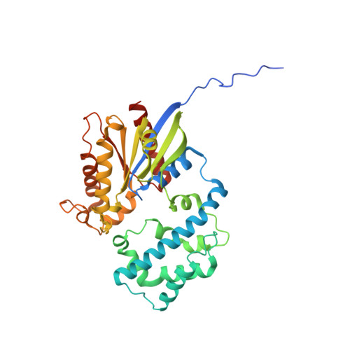
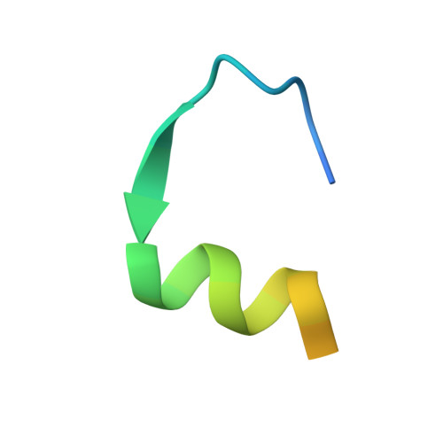
Structural basis for GPCR-independent activation of heterotrimeric Gi proteins.

Kalogriopoulos, N.A.Rees, S.D.Ngo, T.Kopcho, N.J.Ilatovskiy, A.V.Sun, N.Komives, E.A.Chang, G.Ghosh, P.Kufareva, I.

(2019) Proc Natl Acad Sci U S A 116: 16394-16403

  • DOI: https://doi.org/10.1073/pnas.1906658116
  • Primary Citation of Related Structures:  
    6MHE, 6MHF

  • PubMed Abstract: 

    Heterotrimeric G proteins are key molecular switches that control cell behavior. The canonical activation of G proteins by agonist-occupied G protein-coupled receptors (GPCRs) has recently been elucidated from the structural perspective. In contrast, the structural basis for GPCR-independent G protein activation by a novel family of guanine-nucleotide exchange modulators (GEMs) remains unknown. Here, we present a 2.0-Å crystal structure of Gαi in complex with the GEM motif of GIV/Girdin. Nucleotide exchange assays, molecular dynamics simulations, and hydrogen-deuterium exchange experiments demonstrate that GEM binding to the conformational switch II causes structural changes that allosterically propagate to the hydrophobic core of the Gαi GTPase domain. Rearrangement of the hydrophobic core appears to be a common mechanism by which GPCRs and GEMs activate G proteins, although with different efficiency. Atomic-level insights presented here will aid structure-based efforts to selectively target the noncanonical G protein activation.


  • Organizational Affiliation

    Department of Medicine, University of California San Diego, La Jolla, CA 92093.


Macromolecules
Find similar proteins by:  (by identity cutoff)  |  3D Structure
Entity ID: 1
MoleculeChains Sequence LengthOrganismDetailsImage
Guanine nucleotide-binding protein G(k) subunit alpha350Rattus norvegicusMutation(s): 0 
Gene Names: Gnai3Gnai-3
UniProt
Find proteins for P08753 (Rattus norvegicus)
Explore P08753 
Go to UniProtKB:  P08753
Entity Groups  
Sequence Clusters30% Identity50% Identity70% Identity90% Identity95% Identity100% Identity
UniProt GroupP08753
Sequence Annotations
Expand
  • Reference Sequence
Find similar proteins by:  (by identity cutoff)  |  3D Structure
Entity ID: 2
MoleculeChains Sequence LengthOrganismDetailsImage
GirdinB [auth C]31Homo sapiensMutation(s): 0 
UniProt & NIH Common Fund Data Resources
Find proteins for Q3V6T2 (Homo sapiens)
Explore Q3V6T2 
Go to UniProtKB:  Q3V6T2
PHAROS:  Q3V6T2
GTEx:  ENSG00000115355 
Entity Groups  
Sequence Clusters30% Identity50% Identity70% Identity90% Identity95% Identity100% Identity
UniProt GroupQ3V6T2
Sequence Annotations
Expand
  • Reference Sequence
Small Molecules
Ligands 2 Unique
IDChains Name / Formula / InChI Key2D Diagram3D Interactions
GDP
Query on GDP

Download Ideal Coordinates CCD File 
C [auth A]GUANOSINE-5'-DIPHOSPHATE
C10 H15 N5 O11 P2
QGWNDRXFNXRZMB-UUOKFMHZSA-N
GOL
Query on GOL

Download Ideal Coordinates CCD File 
D [auth A]GLYCEROL
C3 H8 O3
PEDCQBHIVMGVHV-UHFFFAOYSA-N
Experimental Data & Validation

Experimental Data

  • Method: X-RAY DIFFRACTION
  • Resolution: 2.00 Å
  • R-Value Free: 0.239 
  • R-Value Work: 0.206 
  • R-Value Observed: 0.208 
  • Space Group: P 43 21 2
Unit Cell:
Length ( Å )Angle ( ˚ )
a = 83.78α = 90
b = 83.78β = 90
c = 141.37γ = 90
Software Package:
Software NamePurpose
PHENIXrefinement
BOSdata collection
MOSFLMdata reduction
Aimlessdata scaling
PHENIXphasing

Structure Validation

View Full Validation Report

Currently 6MHF does not have a validation slider image.


Entry History & Funding Information

Deposition Data


Funding OrganizationLocationGrant Number
National Science Foundation (NSF, United States)United StatesNSF/IOS-1444435

Revision History  (Full details and data files)

  • Version 1.0: 2019-07-31
    Type: Initial release
  • Version 1.1: 2019-08-14
    Changes: Data collection, Database references
  • Version 1.2: 2019-08-28
    Changes: Data collection, Database references
  • Version 1.3: 2019-11-27
    Changes: Author supporting evidence
  • Version 1.4: 2023-10-11
    Changes: Data collection, Database references, Refinement description