6M4L

X-ray crystal structure of the E249Q mutant of alpha-amylase I from Eisenia fetida


Experimental Data Snapshot

  • Method: X-RAY DIFFRACTION
  • Resolution: 1.60 Å
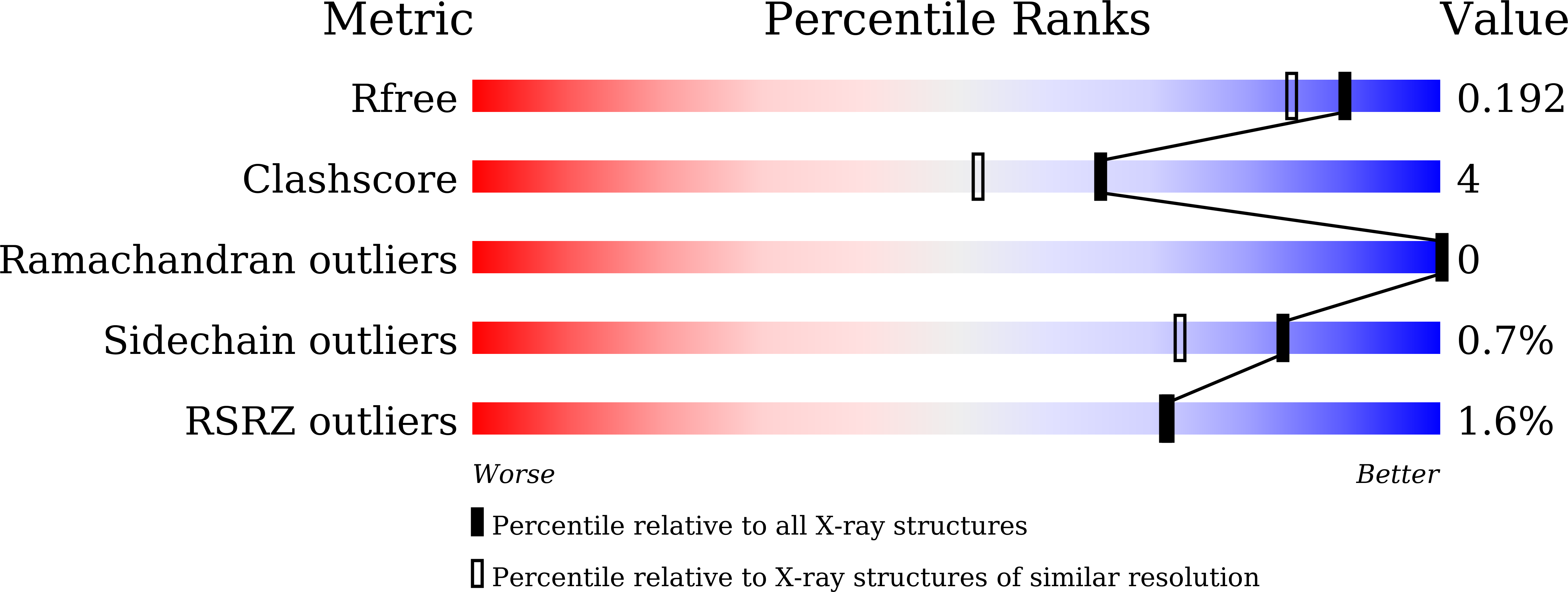
  • R-Value Free: 0.190 
  • R-Value Work: 0.168 
  • R-Value Observed: 0.169 

wwPDB Validation   3D Report Full Report


This is version 1.1 of the entry. See complete history


Literature

X-ray crystallographic structural studies of alpha-amylase I from Eisenia fetida.

Hirano, Y.Tsukamoto, K.Ariki, S.Naka, Y.Ueda, M.Tamada, T.

(2020) Acta Crystallogr D Struct Biol 76: 834-844

  • DOI: https://doi.org/10.1107/S2059798320010165
  • Primary Citation of Related Structures:  
    6M4K, 6M4L, 6M4M

  • PubMed Abstract: 

    The earthworm Eisenia fetida possesses several cold-active enzymes, including α-amylase, β-glucanase and β-mannanase. E. fetida possesses two isoforms of α-amylase (Ef-Amy I and II) to digest raw starch. Ef-Amy I retains its catalytic activity at temperatures below 10°C. To identify the molecular properties of Ef-Amy I, X-ray crystal structures were determined of the wild type and of the inactive E249Q mutant. Ef-Amy I has structural similarities to mammalian α-amylases, including the porcine pancreatic and human pancreatic α-amylases. Structural comparisons of the overall structures as well as of the Ca 2+ -binding sites of Ef-Amy I and the mammalian α-amylases indicate that Ef-Amy I has increased structural flexibility and more solvent-exposed acidic residues. These structural features of Ef-Amy I may contribute to its observed catalytic activity at low temperatures, as many cold-adapted enzymes have similar structural properties. The structure of the substrate complex of the inactive mutant of Ef-Amy I shows that a maltohexaose molecule is bound in the active site and a maltotetraose molecule is bound in the cleft between the N- and C-terminal domains. The recognition of substrate molecules by Ef-Amy I exhibits some differences from that observed in structures of human pancreatic α-amylase. This result provides insights into the structural modulation of the recognition of substrates and inhibitors.


  • Organizational Affiliation

    Institute for Quantum Life Science, National Institutes for Quantum and Radiological Science and Technology, 2-4 Shirakata, Tokai, Ibaraki 319-1106, Japan.


Macromolecules
Find similar proteins by:  (by identity cutoff)  |  3D Structure
Entity ID: 1
MoleculeChains Sequence LengthOrganismDetailsImage
Alpha-amylase520Eisenia fetidaMutation(s): 1 
Gene Names: alpha-amylase
EC: 3.2.1.1
UniProt
Find proteins for A0A173N065 (Eisenia fetida)
Explore A0A173N065 
Go to UniProtKB:  A0A173N065
Entity Groups  
Sequence Clusters30% Identity50% Identity70% Identity90% Identity95% Identity100% Identity
UniProt GroupA0A173N065
Sequence Annotations
Expand
  • Reference Sequence
Small Molecules
Ligands 8 Unique
IDChains Name / Formula / InChI Key2D Diagram3D Interactions
PG4
Query on PG4

Download Ideal Coordinates CCD File 
J [auth A],
O [auth A]
TETRAETHYLENE GLYCOL
C8 H18 O5
UWHCKJMYHZGTIT-UHFFFAOYSA-N
PGE
Query on PGE

Download Ideal Coordinates CCD File 
N [auth A]TRIETHYLENE GLYCOL
C6 H14 O4
ZIBGPFATKBEMQZ-UHFFFAOYSA-N
TRS
Query on TRS

Download Ideal Coordinates CCD File 
I [auth A]2-AMINO-2-HYDROXYMETHYL-PROPANE-1,3-DIOL
C4 H12 N O3
LENZDBCJOHFCAS-UHFFFAOYSA-O
SO4
Query on SO4

Download Ideal Coordinates CCD File 
D [auth A],
E [auth A],
F [auth A]
SULFATE ION
O4 S
QAOWNCQODCNURD-UHFFFAOYSA-L
EDO
Query on EDO

Download Ideal Coordinates CCD File 
K [auth A],
L [auth A],
M [auth A]
1,2-ETHANEDIOL
C2 H6 O2
LYCAIKOWRPUZTN-UHFFFAOYSA-N
ACT
Query on ACT

Download Ideal Coordinates CCD File 
G [auth A],
H [auth A]
ACETATE ION
C2 H3 O2
QTBSBXVTEAMEQO-UHFFFAOYSA-M
CA (Subject of Investigation/LOI)
Query on CA

Download Ideal Coordinates CCD File 
B [auth A]CALCIUM ION
Ca
BHPQYMZQTOCNFJ-UHFFFAOYSA-N
CL (Subject of Investigation/LOI)
Query on CL

Download Ideal Coordinates CCD File 
C [auth A]CHLORIDE ION
Cl
VEXZGXHMUGYJMC-UHFFFAOYSA-M
Experimental Data & Validation

Experimental Data

  • Method: X-RAY DIFFRACTION
  • Resolution: 1.60 Å
  • R-Value Free: 0.190 
  • R-Value Work: 0.168 
  • R-Value Observed: 0.169 
  • Space Group: P 32 2 1
Unit Cell:
Length ( Å )Angle ( ˚ )
a = 96.897α = 90
b = 96.897β = 90
c = 121.957γ = 120
Software Package:
Software NamePurpose
PHENIXrefinement
XDSdata reduction
Aimlessdata scaling
PHASERphasing

Structure Validation

View Full Validation Report


Entry History & Funding Information

Deposition Data


Funding OrganizationLocationGrant Number
Japan Society for the Promotion of Science (JSPS)Japan--

Revision History  (Full details and data files)

  • Version 1.0: 2020-09-16
    Type: Initial release
  • Version 1.1: 2023-11-29
    Changes: Data collection, Database references, Refinement description