6M2P

Crystal structure of a NIR-emitting DNA-stabilized Ag16 nanocluster (A10-deletion mutant)

  • Classification: DNA
  • Organism(s): synthetic construct
  • Mutation(s): No 

  • Deposited: 2020-02-28 Released: 2020-07-01 
  • Deposition Author(s): Kondo, J., Cerretani, C., Vosch, T.
  • Funding Organization(s): Ministry of Education, Culture, Sports, Science and Technology (Japan)

Experimental Data Snapshot

  • Method: X-RAY DIFFRACTION
  • Resolution: 1.13 Å
  • R-Value Free: 0.096 
  • R-Value Work: 0.085 
  • R-Value Observed: 0.086 

wwPDB Validation   3D Report Full Report


This is version 2.1 of the entry. See complete history


Literature

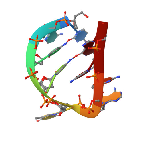
Removal of the A10 adenosine in a DNA-stabilized Ag16 nanocluster.

Cerretani, C.Kondo, J.Vosch, T.

(2020) RSC Adv 10: 23854-23860


Macromolecules

Find similar nucleic acids by:  Sequence   |   3D Structure  

Entity ID: 1
MoleculeChains LengthOrganismImage
DNA (5'-D(*CP*AP*CP*CP*TP*AP*GP*CP*G)-3')
A, B, C, D
9synthetic construct
Sequence Annotations
Expand
  • Reference Sequence
Small Molecules
Ligands 3 Unique
IDChains Name / Formula / InChI Key2D Diagram3D Interactions
AG (Subject of Investigation/LOI)
Query on AG

Download Ideal Coordinates CCD File 
AA [auth C]
BA [auth C]
CA [auth C]
DA [auth C]
E [auth A]
AA [auth C],
BA [auth C],
CA [auth C],
DA [auth C],
E [auth A],
EA [auth C],
F [auth A],
G [auth A],
GA [auth D],
H [auth A],
HA [auth D],
I [auth A],
IA [auth D],
J [auth A],
JA [auth D],
K [auth A],
KA [auth D],
L [auth A],
LA [auth D],
MA [auth D],
NA [auth D],
O [auth B],
P [auth B],
Q [auth B],
R [auth B],
S [auth B],
T [auth B],
U [auth B],
V [auth B],
X [auth C],
Y [auth C],
Z [auth C]
SILVER ION
Ag
FOIXSVOLVBLSDH-UHFFFAOYSA-N
CA
Query on CA

Download Ideal Coordinates CCD File 
M [auth A],
OA [auth D]
CALCIUM ION
Ca
BHPQYMZQTOCNFJ-UHFFFAOYSA-N
CL (Subject of Investigation/LOI)
Query on CL

Download Ideal Coordinates CCD File 
FA [auth C],
N [auth A],
PA [auth D],
W [auth B]
CHLORIDE ION
Cl
VEXZGXHMUGYJMC-UHFFFAOYSA-M
Experimental Data & Validation

Experimental Data

  • Method: X-RAY DIFFRACTION
  • Resolution: 1.13 Å
  • R-Value Free: 0.096 
  • R-Value Work: 0.085 
  • R-Value Observed: 0.086 
  • Space Group: P 1 21 1
Unit Cell:
Length ( Å )Angle ( ˚ )
a = 24.211α = 90
b = 33.713β = 98.11
c = 58.551γ = 90
Software Package:
Software NamePurpose
PHENIXrefinement
XDSdata reduction
XSCALEdata scaling
PHASERphasing
PDB_EXTRACTdata extraction

Structure Validation

View Full Validation Report


Entry History & Funding Information

Deposition Data


Funding OrganizationLocationGrant Number
Ministry of Education, Culture, Sports, Science and Technology (Japan)Japan--

Revision History  (Full details and data files)

  • Version 1.0: 2020-07-01
    Type: Initial release
  • Version 2.0: 2023-03-22
    Type: Coordinate replacement
    Reason: Ligand identity
    Changes: Atomic model, Author supporting evidence, Data collection, Database references, Derived calculations, Non-polymer description, Refinement description, Structure summary
  • Version 2.1: 2023-11-29
    Changes: Data collection, Refinement description