6LUP

Crystal structure of shark MHC CLASS I for 2.3 angstrom


Experimental Data Snapshot

  • Method: X-RAY DIFFRACTION
  • Resolution: 2.30 Å
  • R-Value Free: 0.283 
  • R-Value Work: 0.255 

wwPDB Validation   3D Report Full Report


This is version 1.2 of the entry. See complete history


Literature

The Structure of a Peptide-Loaded Shark MHC Class I Molecule Reveals Features of the Binding between beta 2 -Microglobulin and H Chain Conserved in Evolution.

Wu, Y.Zhang, N.Wei, X.Lu, S.Li, S.Hashimoto, K.Dijkstra, J.M.Xia, C.

(2021) J Immunol 

  • DOI: https://doi.org/10.4049/jimmunol.2001165
  • Primary Citation of Related Structures:  
    6LUO, 6LUP

  • PubMed Abstract: 

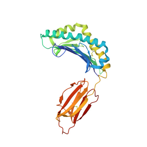
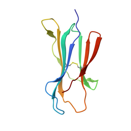
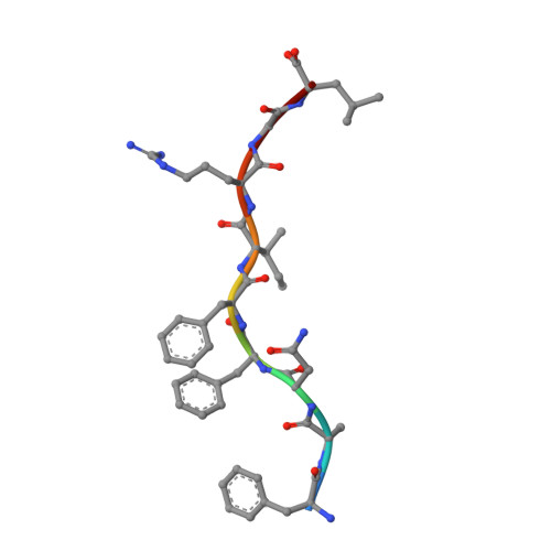
    Cartilaginous fish are the most primitive extant species with MHC molecules. Using the nurse shark, the current study is, to the best of our knowledge, the first to present a peptide-loaded MHC class I (pMHC-I) structure for this class of animals. The overall structure was found to be similar between cartilaginous fish and bony animals, showing remarkable conservation of interactions between the three pMHC-I components H chain, β 2 -microglobulin (β 2 -m), and peptide ligand. In most previous studies, relatively little attention was given to the details of binding between the H chain and β 2 -m, and our study provides important new insights. A pronounced conserved feature involves the insertion of a large β 2 -m F56+W60 hydrophobic knob into a pleat of the β-sheet floor of the H chain α1α2 domain, with the knob being surrounded by conserved residues. Another conserved feature is a hydrogen bond between β 2 -m Y10 and a proline in the α3 domain of the H chain. By alanine substitution analysis, we found that the conserved β 2 -m residues Y10, D53, F56, and W60-each binding the H chain-are required for stable pMHC-I complex formation. For the β 2 -m residues Y10 and F56, such observations have not been reported before. The combined data indicate that for stable pMHC-I complex formation β 2 -m should not only bind the α1α2 domain but also the α3 domain. Knowing the conserved structural features of pMHC-I should be helpful for future elucidations of the mechanisms of pMHC-I complex formation and peptide editing.


  • Organizational Affiliation

    Department of Microbiology and Immunology, College of Veterinary Medicine, China Agricultural University, Beijing, China; and.


Macromolecules
Find similar proteins by:  (by identity cutoff)  |  3D Structure
Entity ID: 1
MoleculeChains Sequence LengthOrganismDetailsImage
MHC class I protein
A, D
267Ginglymostoma cirratumMutation(s): 0 
Gene Names: Gici-UAA01
UniProt
Find proteins for Q9MX69 (Ginglymostoma cirratum)
Explore Q9MX69 
Go to UniProtKB:  Q9MX69
Entity Groups  
Sequence Clusters30% Identity50% Identity70% Identity90% Identity95% Identity100% Identity
UniProt GroupQ9MX69
Sequence Annotations
Expand
  • Reference Sequence
Find similar proteins by:  (by identity cutoff)  |  3D Structure
Entity ID: 2
MoleculeChains Sequence LengthOrganismDetailsImage
Beta-2-microglobulin
B, E
96Ginglymostoma cirratumMutation(s): 0 
Gene Names: B2Mb2m
UniProt
Find proteins for F4ZE04 (Ginglymostoma cirratum)
Explore F4ZE04 
Go to UniProtKB:  F4ZE04
Entity Groups  
Sequence Clusters30% Identity50% Identity70% Identity90% Identity95% Identity100% Identity
UniProt GroupF4ZE04
Sequence Annotations
Expand
  • Reference Sequence

Find similar proteins by:  Sequence   |   3D Structure  

Entity ID: 3
MoleculeChains Sequence LengthOrganismDetailsImage
PHE-ALA-ASN-PHE-PHE-ILE-ARG-GLY-LEU
C, F
9synthetic constructMutation(s): 0 
Sequence Annotations
Expand
  • Reference Sequence
Experimental Data & Validation

Experimental Data

  • Method: X-RAY DIFFRACTION
  • Resolution: 2.30 Å
  • R-Value Free: 0.283 
  • R-Value Work: 0.255 
  • Space Group: P 64
Unit Cell:
Length ( Å )Angle ( ˚ )
a = 125.859α = 90
b = 125.859β = 90
c = 132.473γ = 120
Software Package:
Software NamePurpose
REFMACrefinement
HKL-2000data reduction
HKL-2000data scaling
PHASERphasing

Structure Validation

View Full Validation Report


Entry History & Funding Information

Deposition Data

  • Released Date: 2021-04-28 
  • Deposition Author(s): Wu, Y., Xia, C.

Funding OrganizationLocationGrant Number
National Basic Research Program of China (973 Program)China2013CB835302

Revision History  (Full details and data files)

  • Version 1.0: 2021-04-28
    Type: Initial release
  • Version 1.1: 2021-11-10
    Changes: Database references
  • Version 1.2: 2023-11-29
    Changes: Data collection, Derived calculations, Refinement description