6L59

Crystal structure of the alpha gamma heterodimer of human IDH3 in complex with CIT, Mg and ATP binding at allosteric site and Mg, ATP binding at active site.


Experimental Data Snapshot

  • Method: X-RAY DIFFRACTION
  • Resolution: 2.25 Å
  • R-Value Free: 0.227 
  • R-Value Work: 0.200 
  • R-Value Observed: 0.202 

wwPDB Validation   3D Report Full Report


Ligand Structure Quality Assessment 


This is version 1.1 of the entry. See complete history


Literature

Molecular mechanism of the dual regulatory roles of ATP on the alpha gamma heterodimer of human NAD-dependent isocitrate dehydrogenase.

Sun, P.Bai, T.Ma, T.Ding, J.

(2020) Sci Rep 10: 6225-6225

  • DOI: https://doi.org/10.1038/s41598-020-63425-6
  • Primary Citation of Related Structures:  
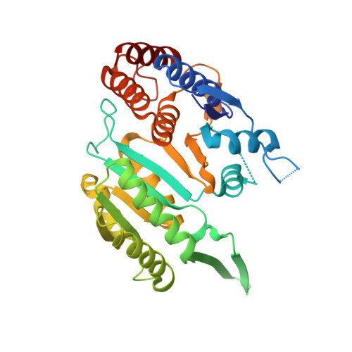
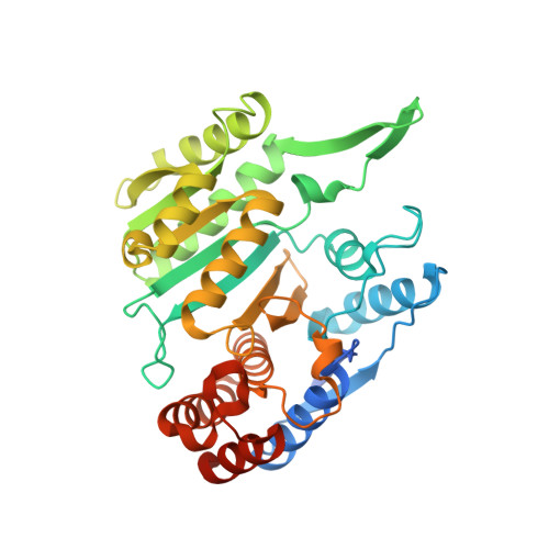
    6L57, 6L59

  • PubMed Abstract: 

    Human NAD-dependent isocitrate dehydrogenase (NAD-IDH) is responsible for the catalytic conversion of isocitrate into α-ketoglutarate in the Krebs cycle. This enzyme exists as the α 2 βγ heterotetramer composed of the αβ and αγ heterodimers. Our previous biochemical data showed that the αγ heterodimer and the holoenzyme can be activated by low concentrations of ATP but inhibited by high concentrations of ATP; however, the molecular mechanism was unknown. Here, we report the crystal structures of the αγ heterodimer with ATP binding only to the allosteric site (α Mg γ Mg+CIT+ATP ) and to both the allosteric site and the active site (α Mg+ATP γ Mg+CIT+ATP ). Structural data show that ATP at low concentrations can mimic ADP to bind to the allosteric site, which stabilizes CIT binding and leads the enzyme to adopt an active conformation, revealing why the enzyme can be activated by low concentrations of ATP. On the other hand, at high concentrations ATP is competitive with NAD for binding to the catalytic site. In addition, our biochemical data show that high concentrations of ATP promote the formation of metal ion-ATP chelates. This reduces the concentration of free metal ion available for the catalytic reaction, and thus further inhibits the enzymatic activity. The combination of these two effects accounts for the inhibition of the enzyme at high concentrations of ATP. Taken together, our structural and biochemical data reveal the molecular mechanism for the dual regulatory roles of ATP on the αγ heterodimer of human NAD-IDH.


  • Organizational Affiliation

    State Key Laboratory of Molecular Biology, CAS Center for Excellence in Molecular Cell Science, Shanghai Institute of Biochemistry and Cell Biology, University of Chinese Academy of Sciences, Chinese Academy of Sciences, 320 Yue-Yang Road, Shanghai, 200031, China.


Macromolecules
Find similar proteins by:  (by identity cutoff)  |  3D Structure
Entity ID: 1
MoleculeChains Sequence LengthOrganismDetailsImage
Isocitrate dehydrogenase [NAD] subunit alpha, mitochondrial342Homo sapiensMutation(s): 0 
Gene Names: IDH3A
EC: 1.1.1.41
UniProt & NIH Common Fund Data Resources
Find proteins for P50213 (Homo sapiens)
Explore P50213 
Go to UniProtKB:  P50213
PHAROS:  P50213
GTEx:  ENSG00000166411 
Entity Groups  
Sequence Clusters30% Identity50% Identity70% Identity90% Identity95% Identity100% Identity
UniProt GroupP50213
Sequence Annotations
Expand
  • Reference Sequence
Find similar proteins by:  (by identity cutoff)  |  3D Structure
Entity ID: 2
MoleculeChains Sequence LengthOrganismDetailsImage
Isocitrate dehydrogenase [NAD] subunit gamma, mitochondrial357Homo sapiensMutation(s): 0 
Gene Names: IDH3G
UniProt & NIH Common Fund Data Resources
Find proteins for P51553 (Homo sapiens)
Explore P51553 
Go to UniProtKB:  P51553
PHAROS:  P51553
GTEx:  ENSG00000067829 
Entity Groups  
Sequence Clusters30% Identity50% Identity70% Identity90% Identity95% Identity100% Identity
UniProt GroupP51553
Sequence Annotations
Expand
  • Reference Sequence
Experimental Data & Validation

Experimental Data

  • Method: X-RAY DIFFRACTION
  • Resolution: 2.25 Å
  • R-Value Free: 0.227 
  • R-Value Work: 0.200 
  • R-Value Observed: 0.202 
  • Space Group: P 31 2 1
Unit Cell:
Length ( Å )Angle ( ˚ )
a = 111.964α = 90
b = 111.964β = 90
c = 145.578γ = 120
Software Package:
Software NamePurpose
HKL-2000data scaling
PHENIXrefinement
PDB_EXTRACTdata extraction
HKL-2000data reduction
PHENIXphasing

Structure Validation

View Full Validation Report


Ligand Structure Quality Assessment 


Entry History & Funding Information

Deposition Data


Funding OrganizationLocationGrant Number
National Natural Science Foundation of ChinaChina31870723 and 31530013

Revision History  (Full details and data files)

  • Version 1.0: 2020-04-29
    Type: Initial release
  • Version 1.1: 2023-11-22
    Changes: Data collection, Database references, Derived calculations, Refinement description